Benign And Malignant Tumors Of Oral Cavity
Question.1. Enumerate white lesions of orofacial region. Describe etiology, histopathology and clinical features of leukoplakia.
Or
Enumerate white lesions of oral cavity. Describe leukoplakia in detail.
Answer. The white lesions of oral cavity/orofacial region are as follows:
- Hereditary condition:
- Leukoedema
- White sponge nevus
- Hereditary benign intraepithelial dyskeratosis
- Keratosis follicularis
- Ptylosis syndrome.
- Leukoplakia and malignancies:
- Chronic cheek biting
- Friction or trauma-associated leukoplakia
- Tobacco associated leukoplakia
- Carcinoma in situ
- Squamous cell carcinoma
- Verrucous carcinoma.
Read And Learn More: Oral Pathology Question And Answers
- Dermatosis:
- Lichen planus
- Lupus erythematous.
- Inflammation:
- Mucous patches of syphilis
- Candidiasis
- Koplik spots of measles.
- Miscellaneous conditions:
- Oral submucous firosis
- Papilloma
- Lipoma
- Hairy tongue
- Geographic tongue
- Fordyce’s granules.
Leukoplakia is defied as “a white patch or plaque that cannot be characterized, clinically or pathologically as any other disease and is not associated with any other physical or chemical causative agent except the use of tobacco”.
Axell et al, 1984 Leukoplakia is defied as a predominantly white lesion of the oral mucosa that cannot be characterized as any other defiable lesion. WHO (1997)
Leukoplakia should be used to recognize white plaques of questionable risk having excluded (other ) known diseases or disorders that carry no increased risk for cancer.
Warnakulasuriya et al (2008).
Etiology Of Leukoplakia
The common predisposing factors of leukoplakia are:
- Tobacco: It is used by large number of people in various forms such as smoking of cigarett, cigar, biddies and pipes.
All these types of tobacco habits are important for development of leukoplakia.
It is believed that during smoking a large amount of tobacco end products are produced in oral cavity.
The products in association with heat cause severe irritation to oral mucus membrane and finally result in development of leukoplakia. - Alcohol: Alcohol leads to the entry of carcinogen into exposed cells and thus alters oral epithelium as well as its metabolism.
- Candidiasis: Chronic candidal infections are associated with leukoplakia.
- Dietary deficiency: Deficiency of vitamin A causes metaplasia and hyperkeratinization of epithelium which may result in development of leukoplakia.
- Syphilis: Syphilitic infections play minor role in causation of leukoplakia.
- Hormonal imbalance: Imbalance or dysfunction of both male and female sex hormones causes keratogenic changes in oral epithelium. These changes lead to the development of leukoplakia.
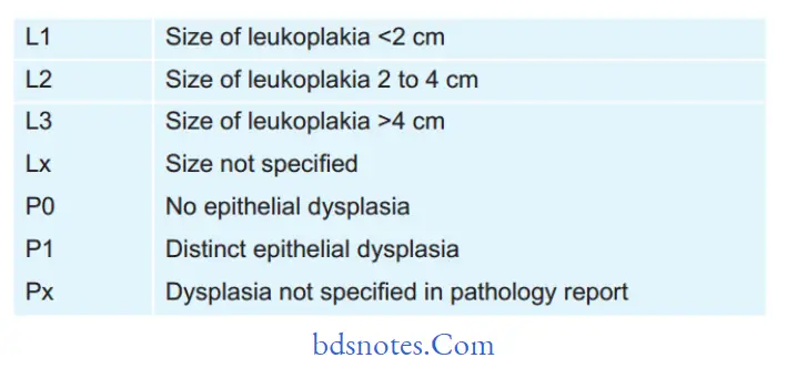
Modifid Classifiation of Leukoplakia
Oral Leukoplakia Staging System
Stage 1 L1P0
Stage 2 L2P0
Stage 3 L3P0 Or L1L2P1
Stage 4 L3P1
Histopathology
During leukoplakia variety of histologic changes are present which are related to:
- Keratinization pattern
- Changes in cellular layer
- Thickness of epithelium
- Alteration in underlying connective tissue stroma.
Keratinization pattern
- Leukoplakia generally presents hyperorthokeratinization or hyperparakeratinization or both with or without the presence of epithelial dysplasia.
- In case of leukoplakia an abnormal increase in the thickness of orthokeratin layer is seen in area of epithelium which is usually keratinized.
- An important histological criterion of leukoplakia is presence of hyperkeratinization of normally keratinized epithelium or some amount of parakeratin deposition in area of epithelium which are usually not keratinized.
- Epithelium dysplasia is more frequently associated with hyperkeratinized lesion.
Changes in Cellular layer
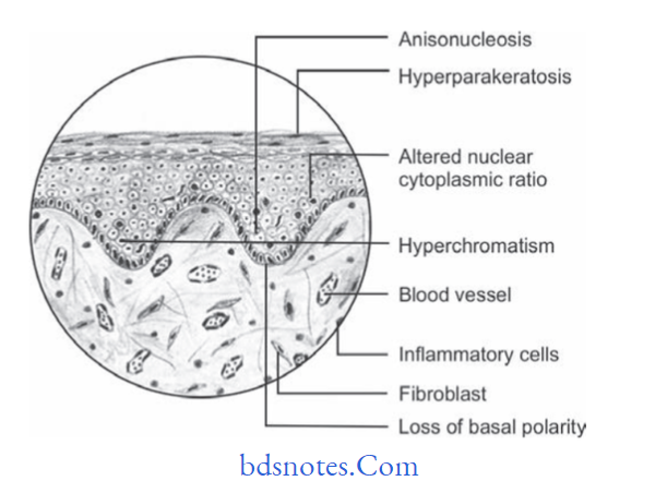
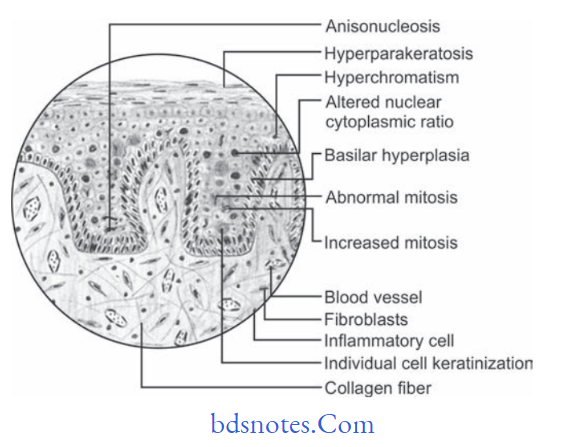
Epithelial dysplasia is the hallmark of histologic changes seen in epithelium in case of leukoplakia. The criteria for epithelial dysplasia are:
Architecture and Cytologic Criteria for Grading Epithelial Dysplasia Given by WHO (2005)
Architecture Criteria
- Irregular epithelial stratifiation
- Loss of polarity of basal cells
- Basal cell hyperplasia
- Drop shaped rete pegs
- Increased number of mitotic fiures
- Abnormally superfiial mitosis
- Dyskeratosis i.e. premature keratinization in the cell
- Keratin pearls within rete ridges
Cytologic Criteria
- Anisonucleosis: Abnormal variation in nuclear size
- Nuclear pleomorphism: Abnormal variation in nuclear shape
- Anisocytosis: Abnormal variation in cell size
- Cellular pleomorphism: Abnormal variation in cell shape
- Increased nuclear cytoplasmic ratio
- Increased nuclear size
- Atypical mitotic fiures
- Increase in the number and size of nucleoli
- Hyperchromatism
Histopathology
- Classification of epithelial dysplasia is done on basis of its severity which is:
- Mild epithelial dysplasia: It refers to the alteration which is limited to basal and parabasal cell layers.
- Moderate epithelial dysplasia: It shows involvement from basal layer to mid portion of spinous cell layer.
- Severe epithelial dysplasia: It shows alterations from basal layer to the level above mid portion of epithelium.
- When complete thickness of epithelium, term carcinoma in situ is used.
- Histopathological report of leukoplakia should include a statement on absence or presence of epithelial dysplasia.
Thickness Of The Epithelium
In leukoplakia, the thickness of epithelium is altered and it occurs in epithelial atrophy or acanthosis.
Alteration In Underlying Connective Tissue
In leukoplakia there is often variable degree of destruction of collagen fiers and moreover chronic inflammatory cell infitrate is also present in underlying connective tissue stroma.
Clinical Features
- Usually the lesion occurs in 4th, 5th, 6th and 7th decades of life.
- Buccal mucosa and commissural areas are most frequent affcted sites followed by alveolar ridge, tongue, hard and soft palate, etc.
- Oral leukoplakia often present solitary or multiple white patches.
- Size of lesion may vary from small well localized patch measuring few millimeters in diameter.
- The surface of lesion may be smooth or fiely wrinkled or even rough on palpation and lesion cannot be removed by scrapping.
- Lesion is whitish or greyish or in some cases it is brownish yellow in color due to heavy use of tobacco.
- In most of the cases these lesion are asymptomatic, however in some cases they may cause pain, feeling of thickness and burning sensation, etc.
Question.2.Define the premalignant lesions and conditions. Describe etiology, histopathology, and clinical features of leukoplakia.
Or
Define and enumerate precancerous lesions. Describe in detail leukoplakia.
Or
Define and enumerate precancerous lesions and conditions. Write in detail about histopathological features of leukoplakia.
Or
Define precancerous lesions and conditions of oral cavity with examples. Describe leukoplakia in detail.
Answer. Premalignant Lesions or Precancerous Lesions
Premalignant lesions are defied as ”A morphologically altered tissue in which cancer is more likely to occur then in its apparently normal counter part”.
The examples are:
- Leukoplakia
- Erythroplakia
- Mucosal changes associated with smoking habits
- Actinic cheilitis, actinic keratosis and actinic elastosis
- Bowen’s disease
- Carcinoma in situ
Premalignant Condition or precancerous Condition
The premalignant condition is defied as “A generalized state of body, which is associated with signifiantly increased risk of cancer development”.
The examples are:
- Oral submucus firosis
- Syphilis
- Lichen planus
- Sideropenic dysphagia
- Lupus erythematosus
- Dyskeratosis congenita
- Xeroderma pigmentosum
- Epidermolysis bullosa.
Question.3. Write notes on hairy leukoplakia.
Answer. Hairy leukoplakia is HIV associated mucosal disorder,which often involves lateral and ventral surfaces of tongue.
Homosexual man with HIV infection may develop white patchy lesion in oral cavity.
Clinical Features
- Clinically hairy leukoplakia occurs more frequently on the lateral border of the tongue however it can also occur on flor of the mouth, buccal mucosa, etc.
- The lesion often appears as white patch and is characterized by an irregular surface, exhibiting numerous linear vertical folds or projections, sometimes so marked to as resemble “Hairs”.
- The lesions are always colonized by Candida albicans.
Hairy leukoplakia probably occurs as an opportunistic infection caused by EpsteinBarr virus. - Hairy leukoplakias are asymptomatic lesions and whenever they occur they occur on buccal mucosa, the lesions are smooth and homogeneous with straitened margin.
Histopathology
- A very characteristic fiding in hairy leukoplakia is presence of subcorneal upper spinous layer zone made up of cytopathologically altered keratinocytes.
- Parakeratin layer is thick often colonized by candidal hyphae.
- The submucosa does not exhibit many inflammatory cell infitrate.
Differential Diagnosis
- Lichen planus
- Verrucous leukoplakia
- Chronic tongue biting habits.
Question.6. Describe etiology, histopathology and clinical features of carcinoma of tongue.
Or
Describe in detail the etiology, clinical features and histopathology of squamous cell carcinoma of tongue.
Answer.
Carcinoma of Tongue Etiology
- Syphilis either an active case or past history of it coexistent with carcinoma of tongue. Syphilis is accepted as having strong association with development of dorsal tongue carcinoma. Arsenicals and heavy metals used to treat syphilis before advent of modern antibiotics have carcinogenic properties themselves and are responsible for some of earlier carcinoma development
in this disease. - Leukoplakia is the common lesion of tongue which leads to carcinoma of tongue.
- Carcinoma of tongue is due to poor oral hygiene, chronic trauma and use of alcohol and tobacco.
- Carcinoma of tongue is due to source of chronic irritation such as carious or broken tooth or an ill fitting denture.
Carcinoma of Tongue Histopathology
- Cells are generally large and show distinct cell membrane.
- Nuclei of neoplastic cells are large.
- Carcinomas of tongue ranges from well diffrentiated keratinizing carcinoma to highly diffrentiated neoplasm.
- Changes of epithelial dysplasia are present.
- There is increased number of mitotic fiure per fild.
Carcinoma of Tongue Clinical Features
- Most common presenting signs of carcinoma of tongue is painless mass or ulcer. The lesion becomes painful when it is secondarily affcted.
- Tumor begins as superfiially indurated ulcer with slightly raised borders and may proceed either to develop a fungating, exophytic mass or to infitrate deep layers of tongue,producing fiation and induration without much surface change.
- If carcinoma is present on the dorsum of tongue,then patient has past or present history of syphilitic glossitis.
- Paresthesia of tongue frequently occurs due to invasion of lingual nerve by tumor cells.
- The common site where the lesion develops is lateral border of tongue and ventral surface of tongue.
- Initial lesions often appear as erythematous macules or nodules or fisured areas over the tongue.
Question.7. Write short note on peripheral giant cell granuloma.
Or
Describe histologic features with diagram of peripheral giant cell granuloma.
Answer. Peripheral giant cell granuloma is the most common of giant cell lesions which arises from tooth bearing areas of jaw and appears as a purplish red nodule.
Peripheral Giant Cell Granuloma Clinical Features
- Lesion usually arises during mixed dentition or during third and fourth decade of life.
- It is most common in males.
- Its site in dentulous patient is interdental papilla. Mandible is more frequently affcted than maxilla.
- Peripheral giant cell granuloma appears as a small, exophytic,well circumscribed, pedunculated lesion on gingival surface.
- Color of lesion varies from purplish red to darkish red.
There can be bleeding from the surface either spontaneously or on provocation from instrument. - Sometimes the peripheral cell granuloma can be aggressive in nature and such lesion may attin very large size and involves some teeth.
In some cases the lesion may develop with an ‘hourglass shape.’
Peripheral Giant Cell Granuloma Histopathology
- Peripheral giant cell granuloma present following histological features.
- Overlying covering epithelium is ulcerated with areas of hemorrhage.
- Underlying connective tissue stroma reveals numerous proliferating firoblasts, blood capillaries and multinucleated giant cells, which are scattred throughout the lesion.
- Fibroblasts present in hypercellular stroma are spindle shaped and have oval shaped nuclei.
- Giant cells are large in size and contain more number of nuclei as compared to true giant cell tumor.
- Areas of hemorrhage and hemosiderin pigment are present within connective tissue stroma.
Peripheral Giant Cell Granuloma Treatment
Surgical excision with curettage.
Question.10. Discuss about central giant cell granuloma.
Answer. Central giant cell granuloma is relatively a common benign intraosseous destructive giant cell lesion which affcts anterior part of jaw bone.
Central Giant Cell Granuloma Clinical Features
- Lesion occurs in young children and female predilection is present.
- Central giant cell granuloma affcts the mandible more often than maxilla and occurs anterior to fist molar.
- Central giant cell granuloma is a small, slow enlarging bony hard swelling of jaw which is painful on palpitation.
- Lesion causes expansion and distortion of cortical plates and there is presence of displacement or mobility of regional teeth.
- Central giant cell granuloma follow an aggressive course and in such cases they produce fast enlarging, large,painful swelling in the jaw.
Central Giant Cell Granuloma Histopathology
- Central giant cell granuloma exhibits fibro vascular connective tissue stroma, consisting of numerous stromal cell which are plum and spindle shaped and undergo frequent mitosis.
- Several areas of hemorrhage and hemosiderin pigmentations are also evident.
- Multiple multinucleated giant cells of varying size are dispersed throughout firous tissue stroma. Giant cells are found around the blood capillaries or near the area of hemorrhage and giant cells consist of 5 to 20 nuclei.
- Small foci of osteoids are often found near periphery of lesion.
- Little amount of chronic inflmmatory cell infitration in connective tissue stroma.
Central Giant Cell Granuloma Radiological Features
- Radiographically the lesion produces multicellular radiolucent area in jaw with soap bubble appearance.
- Margin of lesion is scalloped and well demarcated.
- Resorption of roots of nearby teeth or divergence of roots is common feature.
Central Giant Cell Granuloma Treatment
- Surgical excision is done with curettge.
Question.11.Describe the histopathology of moderately diffrentiated squamous cell carcinoma.
Answer.
Moderately Diffrentiated Squamous Cell Carcinoma Histopathology
- In it the tumor cells are usually more severely dysplastic than of well diffrentiated type.
- Malignant epithelial cells produce little or no keratin and they exhibit greater number of mitotic division.
- Formation of epithelial islands is diminished since tumor cells do not mature properly.
- Malignant tumor cells are recognized as stratifid squamous epithelial cells.
Question.13. Write notes on special stains.
Answer. The special stains are:
Van Gieson’S Stain
- It is a special stain which is used for connective tissue elements. It is used for differentiating between connective tissue fiers and muscle fiers.
- Epithelium (cell and cytoplasm): It takes greenish-yellow stain.
- Collagen fiers are red
- Muscle fiers are yellow
- Nuclei of cells are blue black.
Mallory stain
- It is a special stain for keratin that stains deep orange. It is used in hyperkeratotic lesions.
- The epithelium is royal blue.
- Collagen fiers are royal blue.
- Muscle fiers are royal blue.
- Keratin layers are orange.
- Nucleus is blue black.
Periodic Acid Schiff’S Stain Or Pas Stain
It is a special stain for mucopolysaccharide granules.
These are prominently seen in basement membrane, intercellular spaces and keratin layer.
It is used to detect continuity of basement membrane in intra epithelial carcinoma and squamous cell carcinoma.
- Epithelium and connective tissue is pink.
- Collagen fiers are pink.
- Muscle fiers are pink.
- Nucleus is blue black.
- Granules are of magenta color.
Masson’s trichrome stain
It is a special stain used to diffrentiate between collagen fiers and muscle fiers. It demonstrates connective tissue disorders like leiomyosarcoma and rhabdomyosarcoma.
- Epithelium is red.
- Muscle fiers are bluish violet.
- Collagen fiers and blood vessels are blue.
Question.14.Defie premalignant lesion and condition. Describe etiology, clinical features, and histopathology of oral submucous firosis.
Or
Define precancerous condition and precancerous lesion. Discuss about etiology and histopathology of OSMF.
Answer. The premalignant lesions are defied as morphologically altered tissue in which cancer is more likely to occur then in its apparently normal counter part, e.g. leukoplakia, erythroplakia, nicotiana palati, stomatitis, dyskeratosis congenita, etc.
- The premalignant condition is defied as generalized state of body, which is associated with significantly increased risk of cancer, e.g. oral sub- mucus firosis, syphilis, lichen planus, etc.
Question.15. Enumerate giant cell lesions. Describe in detail central giant cell granuloma.
Answer. Enumeration of giant cell lesions
Giant cell lesions of oral cavity
According To The Nature Of Different Pathologic
Conditions
- Infections
- Bacterial
- Viral
- Fungal
- Protozoal
- Parasitic
- Fibroosseous lesions and osteodystrophies
- Immunologic
- Idiopathic
- Orofacial granulomatosis
- Reaction to materials
- Benign and malignant tumors
In this Classification Giant Cells are Categorized into three Categories
1. Giant cells are the main cause for the pathology
2. Giant cells characterize these lesions
3. Lesions that may be associated with giant cells
Main Cause for the pathology
- Giant cell granuloma
- Peripheral
- Central
- Giant cell tumors
- Giant cell firoma
- Hyperparathyroidism
Giant Cells That Characterize Lesions
- Infections
- Tuberculosis
- Hansen’s disease
- Syphilis
- Measles
- Granulomatous lesions
- Wegener’s granulomatosis
- Orofacial granulomatosis
- Pulse granuloma
- Sarcoidosis
- Lesions in bone
- Aneurysmal bone cyst
- Cherubism
- Paget’s disease
- Foreign body lesions
- Silicosis
- Berylliosis
- Malignancies
- Lymphoma- hodgkin’s disease
- Bronchogenic carcinoma
- Carcinoma of thyroid
- Miscellaneous
- Xanthoma
- Giant cell arteritis
Lesions that may be associated with giant Cell
- Malignancies
- Multiple myeloma
- Ewing’s sarcoma
- Fibrosarcoma
- Chondrosarcoma
- Fibroosseous lesions
- Osteoblastoma
- Fibrous dysplasia
- Cemento-ossifying firoma
- Radicular cyst
Based on their origin
- Epithelially derived, i.e. Warthin-fikeldey giant cells,tumor giant cells
- Stromally derived, i.e. Reedsternberg giant cells.
Question.17. Describe in brief teratoma.
Answer. A true teratoma is a developmental tumor composed of tissues from all three germ layers, i.e. ectoderm, mesoderm and endoderm:
- Such tumors are believed to derive from germ cells or entrapped totipotent blastomeres which can produce derivatives of all three germ layers.
- Teratomas are more common in ovaries and or testis and can be benign or malignant.
Teratoma Clinical Features
- It is most commonly seen in infants.
- Orally it involves soft palate and hard palate. Systemically it involves testis, ovary, abdominal viscera and pineal region.
- It consist of teeth, sebaceous material and some hairs.
Teratoma Histopathology
- H and E-stained section show stratifid squamous epithelium with epithelial appendages consisting of sebaceous glands, sweat glands, hairs, salivary glands, teeth.
- At times thyroid gland tissue and pancreatic tissue can be seen.
Teratoma Treatment
Surgical excision of tumor should be done.
Question.18.Write in detail about Hodgkin’s lymphoma.
Answer. It is also known as hodgkin’s disease.
- Epstein-Barr virus is considered to be the major cause.
- Patients with HIV infection have higher incidence of Hodgkin’s disease.
Hodgkin’s Lymphoma Clinical Features
- Hodgkin’s Lymphoma is most commonly seen in young adults and older individuals.
- More common in males as compared to females.
- Clinical signs and symptoms of the disease are protean
- There is painless enlargement of one or more cervical lymph nodes.
- Palpable painless cervical lymphadenopathy occurs in cervical area, axilla and less commonly in inguinal area and Waldeyers ring and occipital nodes.
- Lymph nodes are fim and rubbery in consistency and overlying skin is normal.
- Symptoms are of unexplained weight loss, fever and night sweats.
Hodgkin’s Lymphoma Histopathology
- Nodular sclerosis hodgkin’s disease
- Morphology show nodular pattrn.
- Broad bands of fiers divide node into nodules
- Characteristic cell is lacunar type ReedSternberg Cell which has monolobated, multilobated nucleus and a small nucleolus with abundant and pale cytoplasm.
- Mixed cellularity hodgkin’s disease
- Infiltrate is usually diffuse
- ReedSternberg cells are of classic type i.e. large with bilobate, double or multiple nuclei and a large eosinophilic inclusion like nucleolus.
- Lymphocyte-depleted hodgkin’s disease
- Infiltrate is diffuse and often appears hypocellular.
- Large number of ReedSternberg cells and bizarre sarcomatous variants are present.
- It is associated with older age and HIV positivity
- Lymphocyte-rich classic hodgkin’s disease
- ReedSternberg cells of the classic or lacunar type are observed with background infitrate of lymphocytes.
- Nodular lymphocyte-predominant hodgkin’s disease
- In this typical ReedSternberg Cell is not seen, instead a variant of Reed
- Sternberg Cell the lymphocytic and histiocytic cells or popcorn cells are seen within the background of inflammatory cells which are predominantly benign lymphocytes.
Hodgkin’s Lymphoma Treatment
Combination of radiotherapy and chemotherapy help in curing the disease.
Question.20.Defie neoplasm. Describe in brief clinical features,X-ray details and histopathology of osteogenic sarcoma.
Answer. “A neoplasm is an abnormal mass of tissue, the growth of which exceeds and is uncoordinated with that of the normal tissues and persists in the same excessive manner after cessation of the stimuli which evoked the change.” Willis
Osteogenic Sarcoma
Osteogenic Sarcoma is a common malignant neoplasm arising from the bone and beside plasma and myeloma.
Osteogenic Sarcoma Clinical Features
- It occurs during 10–25 years of age in the jaw and males are more commonly affected.
- The tumor involves maxilla more often than mandible.
- A very fast enlarging, painful swelling of jaw, causes expansion and distortion of cortical plates.
- Severe facial deformity and diffilty in taking the food due to restricted jaw movement.
- Displacement and loosening of regional teeth.
- Ulceration, hemorrhage and pathological fracture of bone are commonly associated.
Osteogenic Sarcoma X-ray Details
The Xray details of osteosarcoma are divided into following stages, i.e.
Osteogenic Sarcoma Osteolytic Stage
- It reveals moth eaten appearance.
- Border of the lesion at this stage are ill-defied.
- There is perforation and expansion of cortical plates.
- Lamina dura is absent, i.e. it get destroyed.
- Pathological fracture may be present.
- Root resorption is present.
Osteogenic Sarcoma Mixed Stage
- It is called as mixed because this stage show formation and destruction of bone.
- It reveals honeycomb appearance.
- Margins of lesion are ill defied.
Osteogenic Sarcoma Osteoblastic Stage
- It reveals sun ray appearance.
- At times subperiosteal bone laid down in layers which results in onion peel appearance.
- In osteosarcoma periosteum is elevated over the expanding tumor mass in a tent like fashion. At point on the bone where the periosteum begin to merge an acute angle between periosteum and bone is created which is known as
- Codman’s Triangle.
Osteogenic Sarcoma Histopathology
- There will be presence of numerous, actively proliferating,spindle shaped, oval or angular, malignant osteoblast cells within cellular stroma.
- The malignant osteoblast cells exhibit cellular pleomorphism, abnormal increased mitosis and nuclear hyperchromatism
- Multiple areas of newly formed bone or osteoid tissue are often present within firous stroma.
- In chondroblastic variations the malignant tumor cells produce large amount of cartilagenous tissue within tumor.
Question.21.Define oral submucous firosis. Describe in detail etiopathogenesis and histopathology of same.
Answer. OSMF is defined as “An insidious chronic disease affcting any part of oral cavity and sometime pharynx.
Although occasionally preceded by and/or associated with vesicle formation, it is always associated with juxtaepithelial inflammatory reaction followed by firoelastic changes in lamina propria, with epithelial atrophy leading to stiffess of oral mucosa and causing trismus and inability to eat.” — Pindborg (1966)
Oral Submucous Firosis Etiopathogenesis
- Betel nut: Tannic acid and arecoline present in betel nut affct the vascular supply of oral mucosa leading to a neurotropic disorder. Nitrosation of arecoline causes formation of nitrosoguvacine and nitrosoguvacoline as well as 3 methyl nitrosamino propionitrile which leads to alkylation of DNA. Metabolism of nitrosoguvacine and nitrosoguvacoline as well as 3 methyl nitrosamino propionitrile causes formation of cyanoethyl. Cyanoethyl interacts with O’ methyl guanine in DNA. Constant ir
ritation by this leads to OSMF and exposure for long time leads to malignant transformation.
Recent concept: Arecoline in betel nut stimulates fibroblasts. Fibroblasts proliferates and produce collagen. Flavonoids, i.e. catechin and tannin in betel nut stabilizes collagen fiers which make them resistant to degradation by collagenase enzyme. Trismus is caused due to juxtaepithelial hyalinization as well as involvement of the secondary muscles. Increased muscle activity leads to glycogen depletion and reduced blood supply due to connective tissue changes leads to degeneration of muscle and also cause firosis. - Tobacco and lime: Tobacco act as a local irritant which leads to OSMF. Lime causes local irritation as well as vesicle and ulcer formation in the oral mucosa.
- Chilies: Capsaicin is an active ingredient present in red chillies. It is the irritant in chillies which leads to oral submucous firosis.
- Hereditary: Presence of genetic susceptibility is present.
Familial occurrence of OSMF is seen. - Immunological Studies: It was found that human leukocyte antigen (HLA) A10, B7 and DR3 occur signifiantly more frequently in oral submucous firosis.
Question.22.Classify the nonodontogenic tumors of oral cavity and describe firoma.
Or
Write short answer on firoma.
Answer.
Fibroma
Fibroma is a benign tumor of connective tissue origin.
Fibroma Clinical Features
- It is a slow growing lesion and can be seen during 3rd, 4th and 5th decades of life.
- Female predilection is seen.
- It can occur anywhere in the oral cavity but most commonly it is seen on buccal mucosa along plane of occlusion. It also affcts gingiva, tongue, buccal mucosa,lips and palate.
- Lesion appears as elevated nodule of normal color with smooth surface and a sessile or at times pedunculated base.
- Its size can ranges from several millimeters to centimeters.
- Lesion if traumatized it become painful.
- Color of the lesion is pink and texture is smooth.
- Consistency of lesion can be soft or fim or can be elastic.
- At times lesion is traumatized and become inflmed and show ulceration or hyperkeratosis.
Fibroma Histopathology
- Lesion consists of stratifid squamous epithelium which show shortening and fitting of rete pegs.
- Underlying connective tissue stroma show bundles of interlacing collagen fiers which are interspersed with numerous firoblasts.
- There is presence of chronic inflammatory cell infitrate
consisting of lymphocytes and plasma cells. - Areas of calcification and ossifiation can also be seen.
Fibroma Treatment
Excision of the lesion should be done.
Question.23. Enumerate non-odontogenic connective tissue malignant tumors. Discuss in detail about clinical features, etiology and histopathology of firosarcoma.
Answer. Enumeration of non-odontogenic connective tissue malignant tumors
- Fibrous connective tissue: Fibrosarcoma
- Adipose Tissue: Liposarcoma
- Cartilage: Chondrosarcoma
- Bone
- Osteosarcoma
- Osteochondrosarcoma
- Vascular:
- Hemangioendothelioma
- Angiosarcoma
- Kaposi sarcoma
- Neural tissue: Neurosarcoma or Neurofirosarcoma
- Muscle
- Leiomyosarcoma
- Rhabdomyosarcoma
- Lymphoid tissue
- Hodgkin and NonHodgkin lymphoma
- Lymphosarcoma
- Reticular cell sarcoma
- Ewing’s sarcoma
- Burkitts lymphoma
- Multiple myeloma
- Leukemia.
Fibrosarcoma
- Fibrosarcoma is the malignant firous connective tissue tumor and is the malignant tumor of firoblasts.
Fibrosarcoma Clinical Features
- Fibrosarcoma arises at any age but mean age is 40 years.
- Male predilection is seen.
- Fibrosarcoma is most commonly seen in lower extremities i.e. femur and tibia.
- In oral cavity tumor involves mandible, maxilla, maxillary sinus, lip and palate.
- Tumor is generally a large painless mass which lies deep to fascia and has ill-defied margin.
- Associated teeth become mobile.
- Tumors in starting show benign growth and later on they spread rapidly producing large tumor with ulceration and hemorrhage.
- They can also cause pathological fracture.
Fibrosarcoma Etiology
- Most of the firosarcomas arise from preexisting lesions such as Paget’s disease, firous dysplasia chronic osteomyelitis, bone infarcts and in previously irradiated areas of bone.
- Congenital firosarcomas are thought to arise from genetic mutations.
Fibrosarcoma Histopathology
- Various histological grading of firosarcoma are:
Fibrosarcoma Well-differentiated
- In this multiple plump-shaped firoblasts with pale eosinophilic cytoplasm, hyperchromatic spindleshaped nuclei with tapered ends is seen.
- Malignant firoblasts are dispersed in rich collagen area.
- Few mitotic fiures are evident.
Fibrosarcoma Intermediate Grade
- In this tumor show Herring bone pattrn, i.e. parallel sheets of cells arranged in intertwining whorls.
- Cellularity is high.
- Cellular pleomorphism is evident.
- Areas of hyalinization can be appreciated.
Fibrosarcoma High Grade
- Marked cellular atypia and mitotic activity is evident.
- This grade is highly anaplastic and pleomorphic with bizarre nuclei.
Question.24. Write short note on keratoacanthoma.
Answer. Keratoacanthoma is also known as molluscum sebaceum, selfhealing carcinoma or pseudocarcinoma.
Keratoacanthoma clinically and histologically resembles epidermoid carcinoma so it is mistaken as oral carcinoma.
Keratoacanthoma Etiology
- Hereditary predisposition is present.
- Human papilloma virus (HPV) 26 or 37 can lead to keratoacanthoma.
- Sun exposure
- Chemical agents such as coal tar and minerals
Keratoacanthoma Clinical Features
- Its occurrence is at the age of 50 to 70 years. Male predilection is present.
- Intraorally, it is most commonly found on lips.
- Lesion is painful and regional lymphadenopathy is present.
- Lesion is elevated, umblicated with depressed central core with presence of plug of keratin. Lesion appears as dome shaped.
- Margins of lesions are sharply delineated.
- Lesion begins as small nodule which increases in size from 4 to 6 weeks. Later on it undergoes spontaneous regression from 6 to 8 weeks with scar formation.
Keratoacanthoma Histopathological Features
- Hyperplastic squamous epithelium growing into underlying connective tissue.
- Epithelial surface is covered by parakeratin or orthokeratin with parakeratin plugging.
- Pseudocarcinomatous infiltration typically present smooth, regular, well demarcated front which does not extend beyond the level of sweat gland.
- Connective tissue show chronic inflmmatory cell infitration.
- Most characteristic feature of lesion is seen at the margins where normal adjacent epithelium is elevated towards central portion of crater, later on an abrupt change in normal epithelium occurs as hyperplastic acanthotic epithelium is reached.
Keratoacanthoma Treatment
Surgical excision.
Question.25. Write short note on Kaposi’s sarcoma.
Answer. It is also known as multiple idiopathic hemorrhagic sarcoma of Kaposi.
- Kaposi’s sarcoma is the multicentric proliferation of vascular or spindle cell components.
- Kaposi’s sarcoma is currently associated with HIV/AIDS.
Kaposi’s sarcoma Clinical Features
- Clinically, Kaposi’s sarcoma is of four types, i.e.
- Classic (Chronic)
- Endemic (Lymphadenopathic; African)
- Immunosuppression associated (Transplant)
- AIDS related.
Kaposi’s sarcoma Classic type
- Development of cutaneous multifocal blue red nodules on the lower extremities.
- Lesion slowly increases in size and number with some of the lesions extinguishing and new ones forming on adjacent or distinct skin.
- Orally, soft bluish nodules occur on palatal mucosa or gingiva.
Lymphadenopathic
- Present in young African children.
- There is generalized or localized enlargement of lymph node chain which includes cervical lymph nodes.
- Disease follow fulminant course with visceral involvement and minimum skin or mucous membrane involvement.
Immunosuppression Associated
- It is usually seen in renal transplant patients.
- It occurs 1 to 2 years after transplantation
- Progression of disease is directly proportional to loss of cellular immunity of host.
AiDs Related
- Homosexual AIDS patients have maximum chances of developing Kaposi’s sarcoma.
- Lesions occur on cutaneous lesions i.e. along lines of cleavage and tip of nose.
- Oral lesions can occur anywhere in oral cavity, but predilection is for palatal mucosa and gingiva.
- Early oral mucosal lesions are flt and slight blue, red or purple plaques, either focal or diffse and may be completely asymptomatic.
- Later on these lesions may be more deeply discolored and there is development of surface papules and nodules which may become exophytic and ulcerated. These lesions can also bleed.
- Cervical lymphadenopathy and salivary gland enlargement is seen.
Lymphadenopathic Histopathology
Early Lesion Or Patch Stage
- There is proliferation of small veins and capillaries around one or more preexisting dilated vessels.
- Slit like vessels are seen near periphery of preexisting blood vessel, skin adnexa and in between collagen fiers.
- Slit like vessels are lined by plump, mild atypical endothelial cells.
- There is presence of mononuclear inflmmatory cell infitrate consisting of mast cells, scattred erythrocytes and hemosiderin deposit.
Advance Lesion Or Plaque Stage
- There is presence of increased numbers of increased capillaries and dilated vascular channels which are interspersed with proliferating sheets of sarcomatous cells with large number of extravasated erythrocytes.
- Slit like vessels without visible endothelial lining are interspersed with spindle cells.
- Lesional cells show enlarged, hyperchromatic nuclei with mildtomoderate pleomorphism.
Nodular Stage
In all features are more prominent than plaque stage.
Question.26. Write short note on tori.
Answer. Tori are of two types, i.e. torus palatinus and torus mandibularis
Torus Palatinus
Torus Palatinus is a slow growing, flt based boney protuberance which occurs in midline of hard palate.
Torus Palatinus Clinical Features
- Women are affcted more commonly.
- Torus palatinus can occur at any age. It reaches at its peak incidence at age of 30 years.
- Torus Palatinus is an outgrowth in the midline of palate.
- Torus Palatinus is spindle shaped, nodular or lobular.
- Mucosa overlying torus is intact and occasionally it appears blanched.
- Torus Palatinus may become ulcerated if traumatized.
- Torus itself may be composed of dense compact bone or dense compact bone with center of cancellous bone.
Torus Mandibularis
Torus Mandibularis is an exostosis or outgrowth of mandible found on the lingual surface.
Torus Mandibularis Clinical Features
- Growth on lingual surface of mandible occurs at mylohyoid line which is usually opposite to cuspid teeth.
- Mandibular tori are bilateral.
- Bilateral overgrowths are globular or they are multiple.
Torus Mandibularis Treatment
For both tori treatment is surgical excision.
Question.27. Write short note on congenital epulis.
Answer. Congenital Epulis is also known as congenital epulis of newborn or
Neumann’s tumor.
It is benign in nature and mostly occurs as single tumor.
Congenital Epulis Clinical Features
- Tumor is present at birth.
- It is located over maxillary or mandibular gingiva. But more common on maxillary.
- Lesion is pedunculated and is found on incisor region arising from crest of alveolar ridge or alveolar process.
- Lesion varies from few centimeters to millimeters.
Congenital Epulis Histology
- Presence of sheets of large closely packed cells showing fie, granular, eosinophilic cytoplasm which comprises the tumor mass.
- Numerous capillaries are present.
Congenital Epulis Treatment
- Surgical Excision.
Question.28. Enumerate malignant epithelial tumors of oral cavity.
Discuss about etiology, clinical features and histopathology of verrucous carcinoma.
Answer. Enumeration of malignant epithelial tumors of oral cavity
- Squamous cell carcinoma
- Intraepidermoid carcinoma
- Adenocarcinoma
- Metastatic carcinoma
- Basal cell carcinoma
- Transitional cell carcinoma
- Malignant melanoma
- Verrucous carcinoma
- Intraepidermoid carcinoma
- Spindle cell carcinoma
- Primary intra-alveolar carcinoma
- Adenoid squamous cell carcinoma.
verrucous carcinoma Etiology
Tobacco: Verrucous carcinoma develops in tobacco chewers. It usually occurs in the buccal sulcus area where the person holds the tobacco most.
Question.29. Write in brief lipoma.
Answer. Lipoma is a rare intraoral tumor.
Lipoma is a benign, slow growing tumor of mature fat cells.
Lipoma Classification
On the basis of morphology
Superficial: It is a single, yellow, lobulated, painless lesion with sessile or pedunculated base.
Diffse: It occurs in deeper tissues and produces a slight surface elevation.
Encapsulated: It is surrounded by a capsule.
Lipoma Pathogenesis
- HMGIC gene which is mapped to 12q15 is a member of high mobility group protein gene family, play a role in development of lipoma.
- Cells of lipoma are different metabolically as compared to normal fat cells. Precursors of fatt acid should be incorporated at faster rate in fat of lipoma as compared to normal fat and there is reduction in lipoprotein lipase activity.
Lipoma Clinical Features
- Lipomas commonly occur in adults, usually around 3rd and 4th decades of life or in older individuals too.
- Lipomas are soft, smooth surface and nodular masses which can be sessile or pedunculated.
- It is asymptomatic for months or years.
- Buccal mucosa and buccal vestibule are the most common sites which are affected. Other sites involved are tongue,flor of mouth and lips.
- The lesion is 3 cm in size but with time, its size increases up to 5 to 6 cm.
Lipoma Histopathology
- Lesion consists of mature fat cells and can demonstrate a thin firous capsule.
- Lesion shows the lobular arrangement of fat cells.
- Various microscopic variants of lipoma are appreciated histologically, i.e.
- Fibrolipoma: Fibrous component intermixed with lobules of fat cells.
- Angiolipoma: Admixture of mature adipocytes and multiple small blood vessels.
- Spindle cell lipoma: Variable amount of spindle cell in conjunction with lipomatous component.
- Pleomorphic lipoma: Presence of spindle cells along with bizarre,hyperchromatic giant cells.
- Myolipoma: When spindle cells are of smooth muscle origin
- Intramuscular lipomas: Infitrative growth of mature adipocytes which extend between skeletal muscle bundles.
- Myxoid lipoma: If stromal background is myxoid.
Lipoma Treatment
Conservative local excision is the treatment of choice.
Question.30. Write short note on Ewing’s sarcoma—histopathology.
Answer. Following is the histopathology of Ewing’s sarcoma:
- The lesion consists of solid sheets of small round cells with very minimal stroma but at a few places connective tissue septae are present.
- Cells are round and small in shape along with scanty cytoplasm, nuclei of the cell is large round to oval in shape along with dispersed chromatin and hyperchromasia.
- Borders of cell are ill defied.
- Cells also show mitotic fiures.
- Cells of the sarcoma are arranged in fiigree pattrn.
- Small vascular channels are also evident.
- Hemorrhage along with vascular lakes or sinuses are appreciated.
- Perivascular sparing and geographical necrosis is very common in Ewing’s sarcoma.
Question.33. Enumerate the carcinomas of oral cavity. Describe staging and grading of squamous cell carcinoma.
Answer. Enumeration of carcinomas of oral cavity
- Squamous cell carcinoma
- Verrucous carcinoma
- Basaloid squamous cell carcinoma
- Adenoid squamous cell carcinoma
- Spindle cell carcinoma
- Adenosquamous carcinoma
- Undifferentiated carcinoma.
Staging of squamous Cell Carcinoma
Staging is defined as extent of spread of tumor within the body.
Staging of squamous cell carcinoma is done by TNM
classification which was given by American Joint Committee
on Cancer (AJCC)
T is suggestive of primary tumor
N is suggestive of regional lymph nodes
M is suggestive of distant metastasis
T primary tumor
TX Primary tumor cannot be assessed.
T0 No evidence of primary tumor
Tis carcinoma in situ
T1 Tumor 2 cm of less in greatest dimension
T2 Tumor more than 2 cm but not more than 4 cm in greatest dimension
T3 Tumor more than 4 cm in greatest dimension
T4a (Lip) Tumor invades through cortical bone, inferior alveolar nerve, flor of mouth or skin (chin or nose).
T4a (Oral Cavity) Tumor invades through cortical bone, intodeep/extrinsic muscle of tongue (genioglossus, hyoglossus,palatoglossus and styloglossus), maxillary sinus or skin of face.
T4b (lip and oral cavity) Tumor invades masticatory space,pterygoid plates or skull base or encases internal carotid artery
N Regional lymph nodes
NX Regional lymph nodes cannot be assessed
N0 No regional lymph node metastasis
N1 Metastasis in a single ipsilateral lymph node, 3 cm or less in greatest dimension.
N2a Metastasis in a single ipsilateral lymph node, more than3 cm but not more than 6 cm in greatest dimension.
N2b Metastasis in multiple ipsilateral lymph nodes, not more than 6 cm in greatest dimension.
N2c Metastasis in bilateral or contralateral lymph nodes, not more than 6 cm in greatest dimension.
N3 Metastasis in a lymph node more than 6 cm in greatest dimension.
M Distant Metastasis
MX Distant metastasis cannot be assessed
M0 No distant metastasis
M1 Distant metastasis.
Stage Grouping Of Oral Cancer
Grading Of Squamous Cell Carcinoma
- Grading is defined as macroscopic and microscopic degree of differentiation of a tumor.
- Squamous cell carcinoma is divided in following categories by Broder also known as Broder’s classification:
Broader’s Classification
- Grade I Well differentiated, – <25% undiffrentiated cells
- Grade II Moderately differentiated – <50% undifferentiated cells
- Grade III Poorly differentiated – <75% undifferentiated cells
- Grade IV Anaplastic/Pleomorphic – >75% undifferentiated cells
Question.34. Write short note on radiological features of osteosarcoma.
Answer. The radiographic features of osteosarcoma are divided into following stages, i.e.
- Osteolytic Stage
- It reveals moth eaten appearance.
- Border of the lesion at this stage are ill defied.
- There is perforation and expansion of cortical plates.
- Lamina dura is absent, i.e. it get destroyed.
- Pathological fracture may be present.
- Root resorption is present.
- Mixed Stage
- It is called as mixed because this stage shows formation and destruction of bone.
- It reveals honeycomb appearance.
- Margins of lesion are ill defied.
- Osteoblastic Stage
- It reveals sun ray appearance.
- At times subperiosteal bone laid down in layers which results in onion peel appearance.
- In osteosarcoma periosteum is elevated over the expanding tumor mass in a tent like fashion.
At point on the bone where the periosteum begin to merge an acute angle between periosteum and bone is created which is known as Codman’s Triangle.
Leave a Reply