Basal nuclei
Question 1. What are basal nuclei? List their functions.
Answer.
The basal nuclei are large masses of grey matter located in the basal part of the cerebral hemisphere. They include the corpus striatum, claustrum and amygdaloid body.
Basal Nuclei Functions
- To control the automatic associated movements like swinging of arms during walking.
- To help in smoothening the voluntary motor activities of the body.
- To prevent the occurrence of involuntary movements.
Read And Learn More: Selective Anatomy Notes And Question And Answers
Question 2. Write a short note on the corpus striatum.
Answer.
The corpus striatum is situated lateral to the thalamus. Topographically, it is almost completely divided into two parts: the caudate and lentiform nuclei by a band of fibres of the internal capsule. However, anteroinferiorly the two parts are connected with each other by a thin band of grey matter across the anterior limb of the internal capsule.
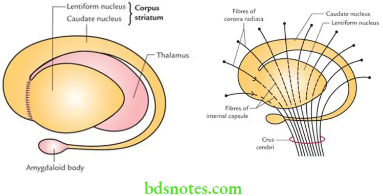
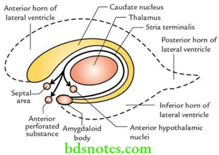
Caudate Nucleus: The caudate nucleus is a large, comma-shaped mass of grey matter that surrounds the thalamus and is itself surrounded by the lateral ventricle. It is divided into three parts – head, body and tail.
- The head is large and rounded and lies in the floor and lateral wall of the anterior horn of the lateral ventricle. Its lower part is connected with the putamen by thin bands of grey matter.
- The body is long and narrow. It lies in the lateral part of the central part of the lateral ventricle.
- The tail is long and slender. It runs forward in the roof of the inferior horn of the lateral ventricle and merges with the amygdaloid nucleus.
Lentiform nucleus: The lentiform nucleus is a large, lens-shaped (biconvex lens) nucleus. In the horizontal section of the cerebrum, it appears wedge-shaped. It has three surfaces: lateral, medial and inferior. Its lateral surface is highly convex.
It is related to the external capsule, which separates it from the claustrum. Its medial surface is also highly convex. It is related to the internal capsule, which separates it from the head of the caudate nucleus and the thalamus. Its inferior surface is related to the sublentiform part of the internal capsule and lies close to the anterior perforated substance.
Question 3. Enumerate the disorders that may occur due to the lesions of basal nuclei.
Answer.
The lesions of basal nuclei lead to various forms of involuntary movements such as:
- Parkinsonism (see below).
- Chorea, choreiform movements as in break dancing.
- Athetosis, athetoid movements, i.e. slow, sinuous and writhing movements.
- Ballismus is a violent burst of irregular movements in the trunk, girdles and limbs.
Question 4. Write a short note on Parkinsonism.
Answer.
Parkinsonism is a degenerative disease involving substantia nigra and/or nigrostriatal fibres causing a deficiency of dopamine in the striatum. The disease usually occurs after 50 years of age.
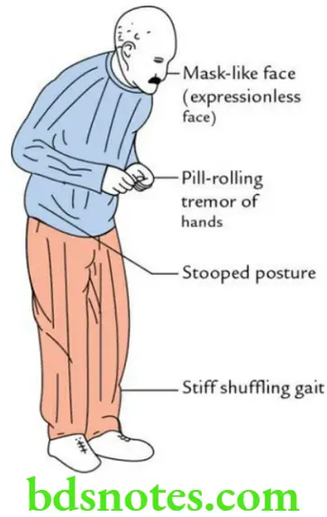
The clinical signs of parkinsonism include:
- Bradykinesia
- Stooped posture
- Shuffling gait
- Cog-wheel rigidity
- Pill-rolling tremors
- Masked facies
- Resting tremors
Limbic system
Question 1. What are the components of hippocampal formation?
Answer.
This hippocampal formation consists of:
- Hippocampus
- Dentate gyrus
- Indusium griseum
- Medial and lateral longitudinal striae
Question 2. Write a short note on the hippocampus.
Answer.
The hippocampus is the area of the cerebral cortex that has rolled on itself in the floor of the inferior horn of the lateral ventricle during fetal life.
It is so named because of its resemblance to ‘sea horse’ in the coronal section.
Histologically, it consists of three layers:
- Superficial polymorphic layer
- The middle pyramidal cell layer
- The deep molecular cell layer
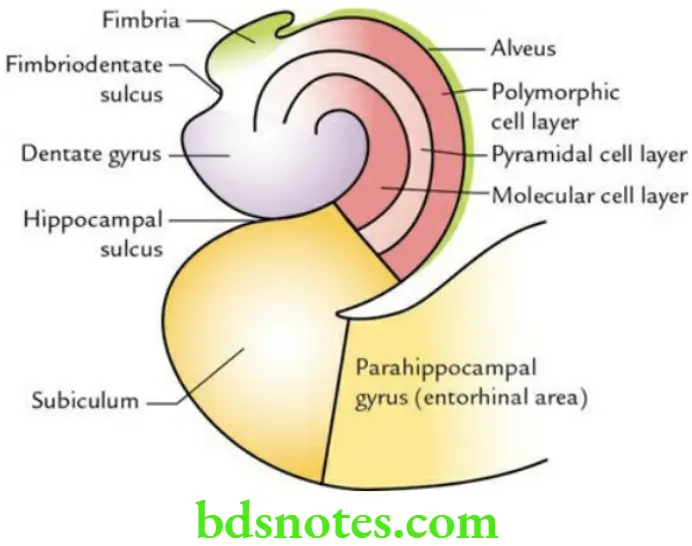
The ventricular surface of the hippocampus is covered by a thin layer of white fibres called alveus. Near the medial border, the fibres of the alveus converge to form the fimbria of the hippocampus.
Hippocampus Function It plays an important role in recent memory.
hippocampus Applied Anatomy The lesions of the hippocampus lead to loss of recent memory (amnesia).
Question 3. Describe the fornix in brief.
Answer.
The fornix is a large bundle of projection fibres (mainly) that connect the hippocampus with the mammillary body. On the medial surface of the cerebral hemisphere, it is seen as an arched bundle of white fibres beneath the corpus callosum.
Fornix Parts, It consists of:
- Fimbriae
- Crura
- Body
- Columns (anterior columns)
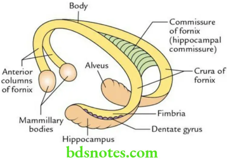
Types of fibres present in the fornix
- Projection fibres, which project from the hippocampus to the mammillary body.
- Commissural fibres, connect the two hippocampi.
- Association fibres, connect the hippocampus with the cingulate gyrus of the same side.
Lateral ventricle
Question 1. Write a short note on the lateral ventricle.
Answer.
- It is a C-shaped cavity within the cerebral hemisphere.
- There are two lateral ventricles, one in each cerebral hemisphere of the cerebrum.
- It is C-shaped and wraps itself around the thalamus, lentiform nucleus and caudate nucleus.
- Each lateral ventricle is situated lateral to the septum pellucidum and below the corpus callosum.
- It is lined by an ependyma and is filled with CSF.
- It has a capacity of about 7–10 mL.
- It communicates with the 3rd ventricle through the interventricular foramen (of Monro).
Lateral Ventricle Parts Each lateral ventricle is divided into four parts:
- Central part/body
- Anterior horn
- Posterior horn
- Inferior horn
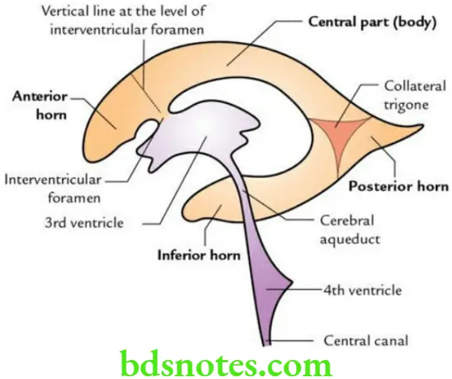
Lateral Ventricle Applied Anatomy The blockage of interventricular foramina leads to excessive accumulation of CSF in the lateral ventricles causing a clinical condition called hydrocephalus.
Question 2. Discuss the boundaries of the central part of the lateral ventricle.
Answer.
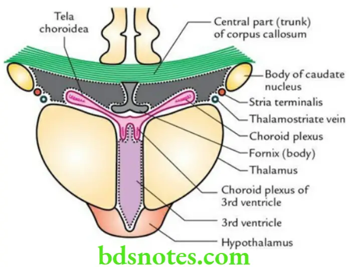
Boundaries It is triangular in shape in a coronal section and presents a roof, floor and medial wall.
Roof: It is formed by the inferior surface of the corpus callosum.
Floor: From lateral to medial side, it is formed by:
- Body of caudate nucleus
- Stria terminalis
- Thalamostriate vein
- The lateral part of the upper surface of the thalamus
- Choroid plexus
- Body of fornix (upper surface)
Medial wall: The medial wall is formed by the septum pellucidum.
Question 3. Discuss the boundaries of the anterior horn of the lateral ventricle.
Answer.
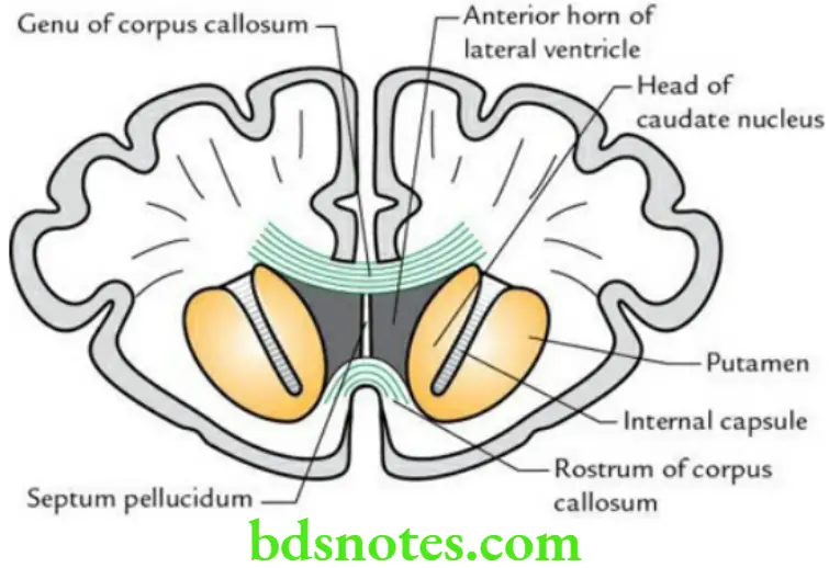
Boundaries It is roughly triangular in coronal section and presents a roof, floor, anterior wall, medial wall and lateral wall.
Roof: It is formed by the anterior part of the body of the corpus callosum.
Floor: It is formed by the upper surface of the rostrum of the corpus callosum.
Anterior wall: It is formed by the genu of the corpus callosum.
Medial wall: It is formed by the septum pellucidum.
Lateral wall: It is formed by the head of the corpus callosum.
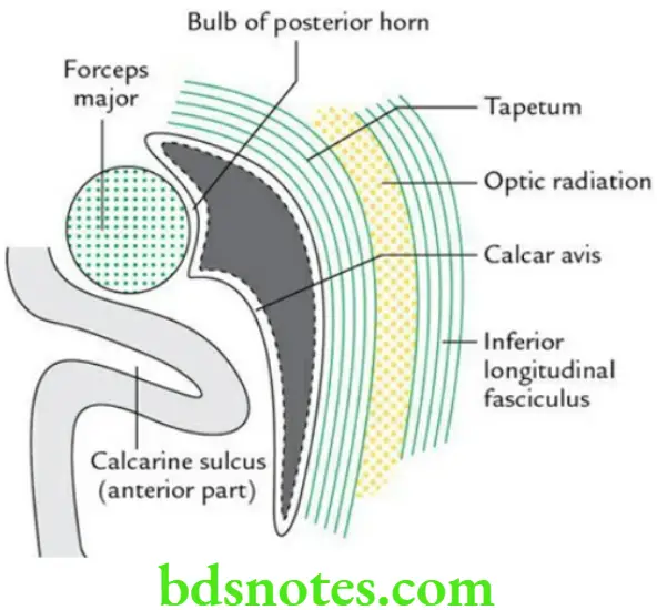
Question 4. Discuss the boundaries of the posterior horn of the lateral ventricle.
Answer.
Boundaries It is quadrangular or diamond-shaped in coronal section and presents a roof, lateral wall, floor and medial wall.
Roof, lateral wall and floor: These are formed by tapetum.
Medial wall: It is formed:
- In the upper part by a bulb of the posterior horn (an elevation/raised area formed by the forceps major).
- In the lower part calcar avis (a raised area formed by the anterior part of calcarine sulcus).
Question 5. Discuss the boundaries of the inferior horn of the lateral ventricle.
Answer.
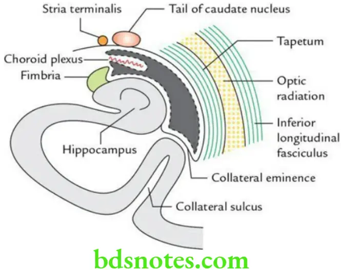
Boundaries It appears on a transverse slit in the coronal section and presents the roof and floor.
Roof: It is formed by:
- Tapetum, covered on its superficial surface by optic radiation and inferior longitudinal fasciculus
- Tail of caudate nucleus
- Stria terminalis
- Amygdaloid body (sometimes)
Floor: From lateral to medial side, it is formed by:
- Collateral eminence, formed by collateral sulcus
- The hippocampus is covered by a thin layer of white matter called the alveus
- Fimbria, formed by alveus
- Choroid plexus

Leave a Reply