Question 1. Axillary nerve
Answer:
- Axillary nerve is one of the two large terminal branches of the posterior cord of branchial plexus
Axillary nerve Course:
- From its origin, it passes backwards between subscapularis & teres major through the quadrangular space
- Here it lies in contact with the surgical neck of the humerus, just below the capsule of the shoulder joint
Read And Learn More: BDS Previous Examination Question And Answers
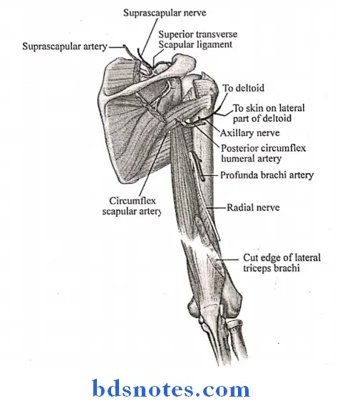
Axillary nerve Branches:
- Muscular branchsupply deltoid & teres minor muscles
- Articular branch-supply shoulder joint
- Cutaneous branchsupply upper lateral cutaneous nerve of arm
- Branch to the shoulder joint which is divides into
- Anterior branch
- Supplies deltoid, pierces muscle & reaches skin
- Posterior branch
- Motor nerve to teres minor
- Cutaneous nerve to the arm
- Supplies posterior fibres of deltoid
- Anterior branch
Axillary nerve Applied Anatomy:
- The nerve may be injured during downward dislocation of shoulder joint
- It may be injured during fracture of surgical neck of humerus
- Injury to the nerve paralyses the deltoid muscle causing flattening of shoulder
- Wrongly injected drugs like penicillin may cause paralysis of the nerve
Question 2. Mention boundaries of suboccipital triangle. Describe the contents of the triangle (or) Suboccipital triangle (or) Suboccipital muscles (or) Contents of suboccipital triangle
Answer:
Suboccipital Triangle:
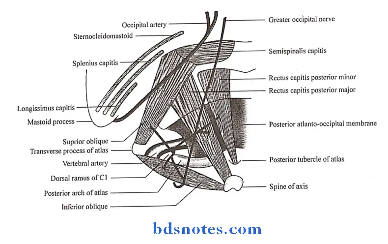
Suboccipital Triangle Boundaries:
1. Superomedially:
- Rectus capitis posterior major
- Rectus capitis posterior minor
2. Superolaterally:
- Superior oblique muscle
3. Inferiorly:
- Inferior oblique muscle
Suboccipital Triangle Roof:
- Medially-semispinalis capitis
- Laterally-longissimus capitis & splenius capitis
Suboccipital Triangle Floor:
- Posterior arch of atlas
- Posterior atlanto-occipital membrane
Suboccipital Triangle Contents:
1. Third part of vertebral artery:
- It appers at the foramen transverium of the atlas, grooves the atlas & leaves the triangle by passing deep to the lateral edge of the posterior atlanto-occipital membrane
- It gives muscular branches to the muscle of the suboccipital region
2. Dorsal rami of C1 nerve:
- It emerges between posterior arch of the atlas & the vertebral artery
- Structures supplied by it are
- Four suboccipital muscles
- Semispinalis capitis
- Nerve to inferior oblique gives off communicating branch to greater occipital nerve
3. Suboccipital plexus of veins:
- It lies in & around the suboccipital triangle & drains
- Muscular veins
- Occipital veins
- Internal vertebral venous plexus
- Condylar emissary vein
4. Suboccipital muscles:
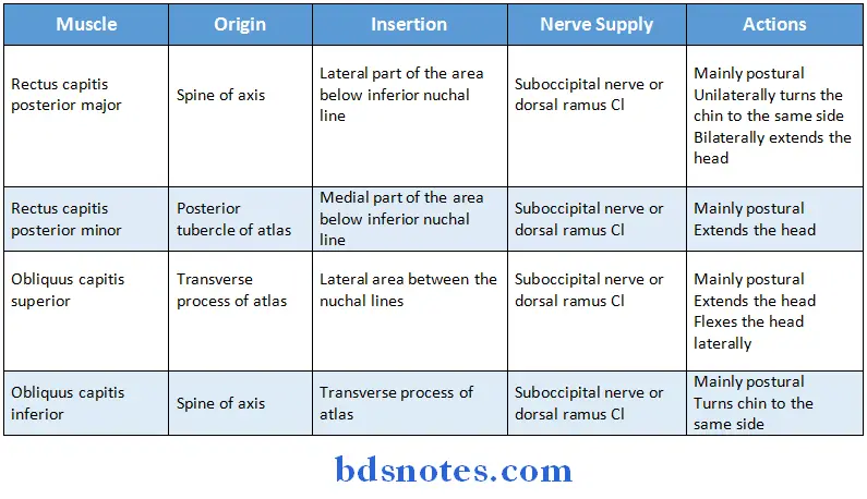
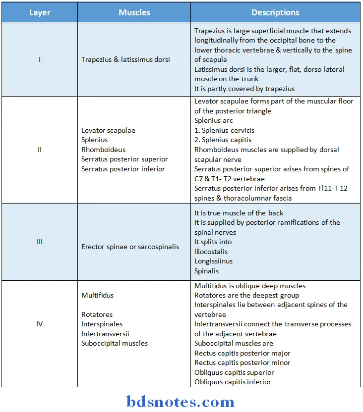
Question 3. Muscles of the back
Answer:
Question 4. Boundaries of Suboccipital Triangle
Answer:
- Superomedially
- Rectus capitis posterior minor
- Rectus capitis posterior major
- Superolaterally
- Superior oblique muscle
- Inferiorly
- Inferior oblique muscle
- Roof
- Medially semispinalis capitis
- Laterally longissimus capitis and splenius capitis
- Floor
- Posterior arch of atlas
- Posterior atlanto-occipital membrane
Question 3. Mention four branches of Cervical Plexus
Answer:
- Superficial branches
- Lesser occipital
- Greater auricular
- Transverse cutaneous nerve of the neck
- Supraclavicular
- Deep branches
- Communicating branches
- Grey rami pass from the superior cervical ganglion to the roots of C1 C1 nerves
- Branch from Ci joins the hypoglossal nerve
- Branch from C2 to the sternocleidomastoid
- Branch from C3 and C, to the trapezius
- Communicating branches
- Muscular branches
- Rectus capitis anterior from C1
- Rectus capitis lateralis from C1, C2
- Longus capitus from C1-C3
- Lower root of ansa cervicalis from C2, C3
Question 5. Vertebral Venous Plexus.
Answer:
- It is made up of a valveless, complicated network of veins
- It anastomoses with superior & inferior venacavae
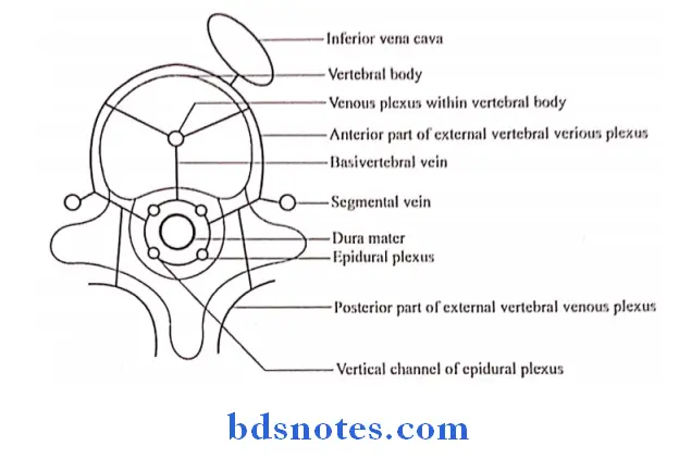
Vertebral Venous Plexus Subdivisions:
1. Epidural plexus
- Lies in the vertebral canal outside the duramater
- It consists of postcentral & prelaminar portion
- It drains the structures in the vertebral canal
- It drains by
- Vertebral veins
- Posterior intercostals veins
- Lumbar veins
- Lateral sacral veins
2. Plexus within the vertebral bodies it drains
- Backwards into the epidural plexus
- Anterolaterally into the external vertebral plexus
3. External vertebral venous plexusconsists of
- Anterior vessels lying in front of vertebral bodies
- Posterior vessels on back of vertebral arches
- It drains by segmental veins
Vertebral Venous Plexus Communications:
- Above with the intracranial venous sinuses
- Below with the pelvic veins, portal veins & caval system of veins
Vertebral Venous Plexus Clinical Anatomy:
- It is the site of development of secondary tumour of breast, the prostrate & the kidney
- Compression of the spinal cord by a tumour gives rise to paraplegia or quadriplegia
- Septic emboli causes collection of pus in the pleural cavity resulting in brain abscess
Question 6. Spinal Duramater.
Answer:
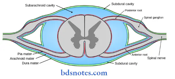
- It is a thick, tough fibrous membrane which forms a loose sheath around the spinal cord
- It is continuous with the meningeal layer of the cerebral duramater
Spinal Duramater Extend:
- From the foramen magnum to the lower border of the second sacral vertebrae
Spinal Duramater Significance:
- It gives tubular prolongations to the dorsal & ventral nerve roots & to the spinal nerves as they pass through the intervertebral foramina
Question 7. Subarachnoid Space.
Answer:
Subarachnoid Space Definition:
- It is a wide intraleptomeningeal space between the piamater & arachnoid space, filled with cerebrospinal fluid
Subarachnoid Space Dimensions:
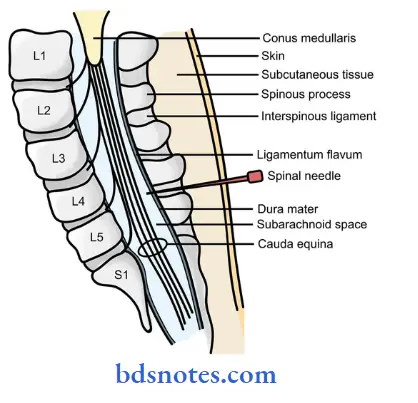
- It is widest below the lower end of the spinal cord where it encloses the cauda equine
Subarachnoid Space Function:
- It surrounds the brain & spinal cord like a water cushion
Subarachnoid Space Clinical Consideration:
- Lumbar puncture is usually done in the lower widest part of the space between 3rd & 4th lumbar vertebrae.
Question 8. Contents of the vertebral canal
Answer:
- Epidural or extradural space
- Thick dura mater
- Subdural capillary space
- Delicate arachnoid mater
- Wide subarachnoid space containing cerebrospinal fluid
- Firm pia mater
- Spinal cord
- Cauda equine
Question 9. Epidural space.
Answer:
Epidural space Location:
- It lies between the spinal dura mater & the periosteum & ligaments lining the vertebral canal
Epidural space Contents:
- Loose areolar tissue
- Semiliquid fat
- Spinal arteries
- It arises from different sources at different levels
- They enter the vertebral canal through the intervertebral foramina
- They supply the spinal cord, the spinal nerve roots, the meninges, the periosteum & ligaments
- The internal vertebral venous plexus
-
- Venous blood from the spinal cord drains into the epidural or internal vertebral plexus
Question 10. Spinal nerves
Answer:
- The spinal cord gives rise to 31 pairs of spinal nerves
- 8 cervical
- 12 thoracic
- 5 lumbar
- 5 sacral
- 1 coccygeal
- Each nerve is attached to the cord by
- Ventral motor root
- Dorsal sensory rootit bears a ganglion
- Both roots unite in the intervertebral foramen to form the nerve trunk
- The root trunk divides into
- Ventral rami
- Dorsal rami
- The roots of spinal nerves are surrounded by sheaths derived from the meninges
- The dural sheath encloses the terminal parts of the roots, continues over the nerve trunk & is lost by merging with the epineurium of the nerve
Question 11. Contents of the intervertebral foramen
Answer:
- The intervertebral foramina are a pair of foramina between the pedicles of the adjacent vertebrae
Intervertebral Foramen Contents:
- The ends of the nerve roots
- The dorsal root ganglion
- The nerve trunk
- The beginning of the dorsal & ventral rami
- A spinal artery
- An intervertebral vein
Question 12. Lumbar puncture
Answer:
Lumbar puncture Procedure:
- Make the patient lying on side with maximally flexed spine
- A line is drawn between highest points of iliac spine at L4 level
- Skin is locally anaesthetized
- Lumbur puncture needle is inserted
Lumbar puncture Site of Injection:
- In adults:
- Between 13 & 14 spines
- In infantsduring 2nd month of life
- Between 14 & 15 spine
Lumbar puncture Penetration of needle:
- Needle is penetrated to the following structures to release the CSP
- Skin fat
- Supraspinous & interspinous ligaments
- Ligamentum flava
- Epidural space
- Dura
- Arachnoid
- Subarachnoid space

Leave a Reply