Question 1. Development of tongue.
Answer:
- Development of the tongue starts in the fourth week of intrauterine life
- The medial most parts of the mandibular arches proliferate to form two lingual swellings.
- These lingual swellings are partially separated from each other by another swelling called tuberculum impair.
- These are derived from 1st branchial arch.
Read And Learn More: BDS Previous Examination Question And Answers
- Another midline swelling, hypobranchial eminence is derived from mesoderm of the second, third, and part of the fourth arch.

- It is sub-divided into cranial and caudal part.
- The anterior two-thirds of the tongue is formed by fusion of
- The tuberculum impair and
- The two lingual swelling.
- The posterior one-third of the tongue is derived from the cranial part of the hypobranchial eminence.
- The posterior-most part of the tongue is derived from the fourth arch.
- The musculature of the tongue is derived from the occipital myotomes.
- Taste buds are formed in relation to the terminal branches of the innervating nerve fibres.
- Nerve supply of the tongue depends on the its developmental derivation.
- Anterior two-third of the tongue is supplied by lingual branch of mandibular nerve which i branchial arch.
- Posterior one third is supplied by the glossopharyngeal nerve, nerve of third branchial arch.
- Posterior most part is supplied by the superior laryngeal nerve, nerve of the fourth branchial arch.
Question 2. Give a detailed account of development of milk teeth.
Answer:

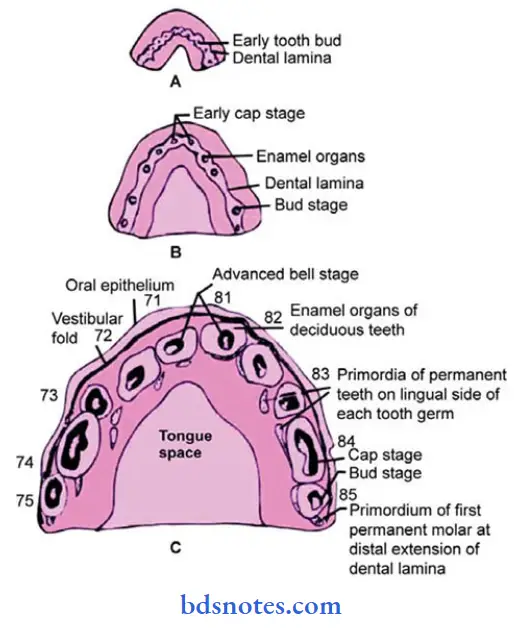
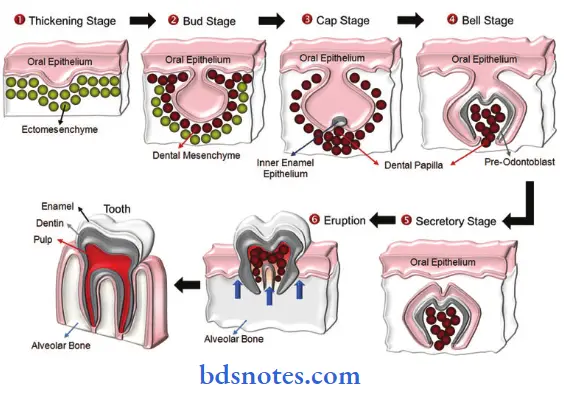
- By the sixth week of development of basal layer of the epithelial lining of the oral cavity forms dental
lamina. - This lamina gives rise to 16 localized thickened buds called enamel organs.
- The enamel organ grows downwards assuming a cup-shaped appearance which is occupied by dental papilla.
- This stage is described as cap stage.
- The cells of the enamel organ lining the papilla become columnar called ameloblasts.
- Mesodermal cells of the papilla form a continuous epithelium-like layer called odontoblast.
- The remaining cells form the pulp.
- The developing tooth now looks like a bell.
- Amelobasts lay down enamel while odontoblasts lay down dentin.
- Odontoblasts in the root region lay down dentin.
- The dentin is covered by mesenchymal which lay down cementum.
- Outside it, mesenchymal cells from the periodontal ligament.
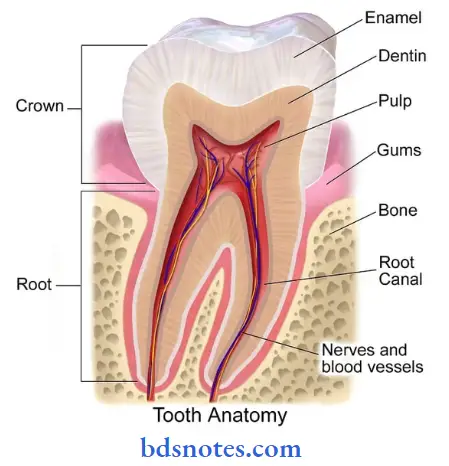
Question 3. Structure of tooth (labelled diagram only)
Answer:

Question 3. Enumerate congenital anomalies of teeth.
Answer:
- Anodontia – absence of teeth.
- Supernumerary teeth – extra number of teeth.
- Abnormal teeth.
- Gemination – fusion of two/more teeth.
- Precocious – early eruption of teeth.
- Delayed eruption of teeth.
- Ectopic eruption of teeth.
- Improper formation of enamel and dentin.
Question 4. Development of oral mucosa.
Answer:
- The oral cavity is derived partly from the ectoderm and partly from the endoderm.
- The anterior part of the oral cavity is derived from the ectoderm while tongue, epiglottis and pharynx are derived from the endoderm.
- The vestibular lamina separates from the primary epithelial band at about 6 weeks.
- Degeneration of the cells in the central part of this process leads to the formation of labial and buccal sulcus.
- The tongue is separated from the rest of the mandibular process by linguo-gingival sulcus.
- The alveolar process of the upper jaw is separated from the upper lip and cheek by labio-gingival sulcus appears laterally to the linguo-gingival sulcus.

Leave a Reply