Question.1. Write short note on Acute Rheumatic Fever.
Or
Describe in brief about rheumatic fever.
Or
Write short answer on rheumatic fever.
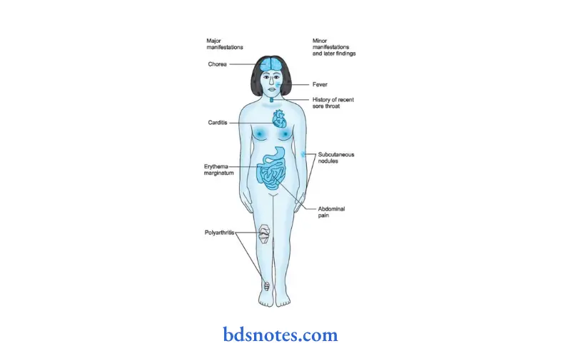
Answer. It is an acute inflmmatory disease which occurs due to infection by group A hemolytic streptococci which involves heart, joint, skin and nervous system which develops as autoimmune reaction to infecting organism.
Acute Rheumatic Fever Etiology
- Predisposing causes:
Age should be 5 to 15 years.
Sex has equal incidence - Genetic factors: Family incidence known.
- Social and economic factors: Dampness, overcrowding and under nutrition increases incidence.
- Idiosyncrasy is presumably a factor since 3% of people are involved in streptococcal epidemics develop rheumatic fever.
Read And Learn More: General Medicine Question And Answers
Clinical Manifestations Acute Rheumatic Fever.
- Prodromal phase: Tonsillitis or sore throat 1 to 4 weeks prior to onset of acute rheumatic fever. Besides this anorexia, pallor, fatigability and nervous irritability is present.
- Latent phase: When antibodies to preceding streptococcal infection are produced.
- Phase of onset of rheumatic fever/mode of onset.
Arthritis and fever 2–3 weeks after infection. - Cardiac symptoms 3–6 weeks after infection is fist to draw attntion.
- Abdominal symptoms: Abdominal pain and tenderness, nausea, vomiting, fever and leukocytosis.
- Pyrexia of unknown origin
- Typhoid or inflenzal mode of onset with fever
- Nodules of skin lesion.
Treatment of acute rheumatic Fever
- Bed rest is important to reduce joint pain and cardiac workload.
Duration of bed rest is guided by markers of inflmmation like temperature, WBC count and ESR. - Benzathine penicillin 1.2 mu IM 4 hourly.
If patient is allergic to penicillin, erythromycin 40–50 mg / kg for ten days is given. - Aspirin usually relieves symptom of arthritis rapidly.
A starting dose of 60 mg / kg body weight per day is given divided into 6 doses.
The dose may be increased to 120 mg / kg body weight.
This dose may produce severe symptoms like vomiting, tachypnea and acidosis is given till ESR comes to normal. - Corticosteroids like prednisolone produces rapid symptomatic relief than aspirin and is indicated in cases with severe arthritis or carditis.
Prednisolone is given in doses of 1.2 mg / kg body weight till ESR comes to normal.
Question.2. Outline the management of acute rheumatic fever.
Or
Discuss the management of acute rheumatic fever.
Answer.
Management Acute Rheumatic Fever.
1. Treatment of acute attck: Rheumatic Fever
- Bed rest is important to reduce joint pain and cardiac workload.
Duration of bed rest is guided by markers of inflmmation like temperature,
WBC count and ESR. - Benzathine penicillin 1.2 mu IM 4 hourly. If patient is allergic to penicillin, erythromycin 40–50 mg / kg for ten days is given.
- Aspirin usually relieves symptom of arthritis rapidly.
A starting dose of60 mg/kg body weight per day is given divided into 6 doses.
The dose may be increased to 120 mg / kg body weight.
This dose may produce severe symptoms like vomiting, tachypnea and acidosis. Aspirin is given till ESR comes to normal. - Corticosteroids like prednisolone produces rapid symptomatic relief than aspirin and is indicated in cases with severe arthritis or carditis.
Prednisolone is given in doses of 1.2 mg / kg body weight till ESR comes to normal
2. Secondary prevention: Rheumatic Fever
To prevent further attck of rheumatic fever, longterm prophylaxis is needed.
- Benzathine penicillin 1.2 mu IM is injected at the interval of 21 days.
Further attck is unusual after the age of 21 years and treatment can be stopped. - To prevent chances of endocarditis prophylactic antibiotic therapy should be given.
Question.3.Describe briefly diagnosis of rheumatic fever.
Or
Write short notes on Jones criteria for rheumatic fever.
Or
Write short note on duke jones criteria in acute rheumatic fever.
Answer. Diagnosis of rheumatic fever is made by ‘Jones criteria’ which is as follows:
Major criteria Rheumatic Fever
1. Carditis: Rheumatic Fever
- It is pancarditis involving endocardium, myocardium and pericardium.
- It manifests as breathlessness, palpitation and chest pain.
- Tachycardia, cardiomegaly and new or change murmurs
- Aortic regurgitation in 50% cases.
- Pericarditis produces frictional rub and pericardial tenderness.
- Cardiac failure due to myocardial infarction.
2. Sydenham’s chorea: Rheumatic Fever
- Late neurological manifestations that occurs at least three months after the episode of acute rheumatic fever when all signs disappear.
- More common in female.
- It is characterized by involuntary dancing movements of hands, feet or face.
3. Polyarthritis: Rheumatic Fever
- Early feature of illness is non-specifi.
- It is characterized by acute painful symmetric and migratory inflmmation of large joints.
- Classical presentation is acute migratory polyarthritis.
Pain and swelling in involved joints subside or disappear as newer joints get affcted.
4. Erythema marginatum: Rheumatic Fever
Red macules which fade in center, but remain red at the edges and occur mainly on trunk and proximal extremities on face.
5. Subcutaneous nodules: Rheumatic Fever
They are small, dense, firm, painless and are best felt over tendons and bones.
- Nodules appear more than 3 weeks after onset ofother manifestations.

Clinical Rheumatic Fever
- Fever
- Arthralgia
- Previous history of rheumatic fever or rheumatic heart disease.
Laboratory Rheumatic Fever
- Acute phase reactants (leucokytosis, raised ESR, C reactive protein)
- Prolonged PR interval in ECG.
Essential criteria Rheumatic Fever
Evidence for recent streptococcal infection as evidenced by:
1. Increase in ASO titer
- > 333 Todd units (in children).
- > 250 Todd units (in adults).
- Positive throat culture for streptococcal infection
- Recent history of scarlet fever.
Confirmation of Diagnosis Rheumatic Fever
Result is based on Presence of two or more major criterias or one major and two minor criteria, in the presence of essential criteria, is required to diagnose acute rheumatic fever.
Question.4. How will you diagnose and manage a case of rheumatic fever? Outline complications of rheumatic fever.
Answer.
Diagnosis of rheumatic fever is made by ‘Jones criteria’ which is as follows:
Major criteria Rheumatic Fever
1. Carditis: Rheumatic Fever
- It is pancarditis involving endocardium, myocardium and pericardium.
- It manifests as breathlessness, palpitation and chest pain.
- Tachycardia, cardiomegaly and new or change murmurs
- Aortic regurgitation in 50% cases.
- Pericarditis produces frictional rub and pericardial tenderness.
- Cardiac failure due to myocardial infarction.
2. Sydenham’s chorea: Rheumatic Fever
- Late neurological manifestations that occurs at least three months after the episode of acute rheumatic fever when all signs disappear.
- More common in female.
- It is characterized by involuntary dancing movements of hands, feet or face.
3. Polyarthritis: Rheumatic Fever
- Early feature of illness is non-specifi.
- It is characterized by acute painful symmetric and migratory inflmmation of large joints.
- Classical presentation is acute migratory polyarthritis.
Pain and swelling in involved joints subside or disappear as newer joints get affcted.
4. Erythema marginatum: Rheumatic Fever
Red macules which fade in center, but remain red at the edges and occur mainly on trunk and proximal extremities on face.
5. Subcutaneous nodules: Rheumatic Fever
They are small, dense, firm, painless and are best felt over tendons and bones.
- Nodules appear more than 3 weeks after onset ofother manifestations.

Clinical Rheumatic Fever
- Fever
- Arthralgia
- Previous history of rheumatic fever or rheumatic heart disease.
Laboratory Rheumatic Fever
- Acute phase reactants (leucokytosis, raised ESR, C reactive protein)
- Prolonged PR interval in ECG.
Essential criteria Rheumatic Fever
Evidence for recent streptococcal infection as evidenced by:
1. Increase in ASO titer
- > 333 Todd units (in children).
- > 250 Todd units (in adults).
- Positive throat culture for streptococcal infection
- Recent history of scarlet fever.
Confirmation of Diagnosis Rheumatic Fever
Result is based on Presence of two or more major criterias or one major and two minor criteria, in the presence of essential criteria, is required to diagnose acute rheumatic fever.
Management Rheumatic Fever
1. Treatment ofacute attck:
- Bed rest is important to reduce joint pain and cardiac workload.
Duration of bed rest is guided by markers of inflmmation like temperature,
WBC count and ESR. - Benzathine penicillin 1.2 mu IM 4 hourly. If patient is allergic to penicillin, erythromycin 40–50 mg / kg for ten days is given.
- Aspirin usually relieves symptom of arthritis rapidly.
A starting dose of60 mg/kg body weight per day is given divided into 6 doses.
The dose may be increased to 120 mg / kg body weight.
This dose may produce severe symptoms like vomiting, tachypnea and acidosis. Aspirin is given till ESR comes to normal. - Corticosteroids like prednisolone produces rapid symptomatic relief than aspirin and is indicated in cases with severe arthritis or carditis.
Prednisolone is given in doses of 1.2 mg / kg body weight till ESR comes to normal
2. Secondary prevention: To prevent further attck of rheumatic fever, longterm prophylaxis is needed.
- Benzathine penicillin 1.2 mu IM is injected at the interval of 21 days.
Further attck is unusual after the age of 21 years and treatment can be stopped. - To prevent chances of endocarditis prophylactic antibiotic therapy should be given.
Complications Rheumatic Fever
- Myocardial infarction
- Mitral stenosis
- Tricuspid regurgitation
- Aortic regurgitation
- Aortic stenosis is rare
- Mitral regurgitation.
Question.5. Enumerate the causes of Jones criteria of acute rheumatic fever.
Answer. The causes of Jones criteria are:
- Previous streptococcal infection
- Recent scarlet fever
- Positive throat culture from streptococcal A
- Increased-antistreptolysin O titer.
Question.6. Describe clinical features, diagnosis, investigations and management of rheumatic mitral stenosis.
Answer. Mitral stenosis is a valvular heart disease.
Rheumatic mitral stenosis occurs in elderly people and is most common in females.
Clinical Manifestations Rheumatic Mitral Stenosis.
Symptoms Rheumatic Mitral Stenosis.
- Patient complains of breathlessness and fatigue on exertion.
- Progression of stenosis lead to dyspnea on rest and even have orthopnea and paroxysmal nocturnal dyspnea.
- Acute pulmonary edema can also occur.
- Hemoptysis can be present due to rupture of pulmonary congestion and pulmonary embolism and cough due to pulmonary congestion.
- Chest pain is present due to pulmonary venous hypertension.
Signs Rheumatic Mitral Stenosis.
- Atrial firillation is present.
- Auscultation: Presence of loud fist heart sound, opening snap and mid diastolic low pitched rumbling murmur best heared at the apex.
- Signs ofraised pulmonary capillary pressure: Pleural effsion, crepitation, pulmonary edema.
- Signs ofpulmonary hypertension: RV heave, loud P2
- Others: Basal crackels, ascites and pleural effsion
Investigations Rheumatic Mitral Stenosis.
1. ECG:
- Right ventricular hypertrophy
- Left atrial hypertrophy
2. X-ray chest:
- Prominent left atrial appendage may be seen in left border of heart between pulmonary artery and left ventricle. It indicates left atrial enlargement.
- Double shadow of enlarged left atrium on right sideof spine.
- Signs of pulmonary venous congestion
3. Echocardiogram:
- Show thick immobile mitral cusp
- Decreased diastolic filing of left ventricle
- Decreased valve orifie area
- Left atrial thrombus, if it is present.
4. Cardiac catheterization: is used to assess valvular lesions
and to detect coronary artery disease.
5. Doppler:
- Pressure gradient across mitral valve
- Pulmonary artery pressure
- Left ventricular function
Diagnosis Rheumatic Mitral Stenosis.
It is based on physical signs and investigations.
Management Rheumatic Mitral Stenosis.
Medicinal: Rheumatic Mitral Stenosis.
- Salt restriction should be done in diet or very low salt diet is given.
- Digitalis therapy is given. In the patient with congestive heart failure Tab. digoxin 0.25 mg BD is given.
- Diuretics can be given for controlling heart failure
- Anticoagulants such as heparin can be given to prevent embolism
- Prophylactic oral penicillin V 250 mg BD is given to prevent rheumatic fever. If patient is allergic of penicillin erythromycin 250 mg daily orally is given.
Surgical: Rheumatic Mitral Stenosis.
When patient remains symptomatic despite of medical treatment or when mitral stenosis is severe, surgical intervention is needed:
1. Mitral valvotomy:
- Percutaneous balloon valvotomy is indicated when mitral valve is noncalcifid and without regurgitation.
The procedure involves the passing of catheter across the valve and infltion of the balloon to dilate the orifie. - Open valvotomy is carried out in patients where balloon valvotomy is not possible or in cases with restenosis.
In this procedure, the fusion of the valve is loosened and calcium deposit and thrombi are removed.
2. Mitral valve replacement: The mitral valve is replaced when there is critical mitral stenosis and/or there is associated mitral regurgitation.
Replacement is also done when the mitral valve is severely distorted and calcifid.
Question.7. Describe briefly clinical features and management of aortic regurgitation.
Answer. Aortic regurgitation is produced due to acute rheumatic carditis which is associated with other valve involvement and infective endocarditis.
Clinical Features Rheumatic Mitral Stenosis.
Symptoms Rheumatic Mitral Stenosis.
1. In mild to moderate aortic regurgitation:
- Often asymptomatic
- On palpitation — pounding of heart is a common symptom
- Symptoms of left heart failure appear but late
2. In severe aortic regurgitation:
- Symptoms of heart failure, i.e., dyspnea, orthopnea are present at onset.
- Angina pectoris is frequent complaint.
- Arrhythmias are uncommon.
Signs Rheumatic Mitral Stenosis.
- Collapsing or good volume pulse (wide pulse pressure)
- Bounding peripheral pulses
- Dancing carotids (Corrigan’s sign)
- Capillary pulsation in nail beds (Quincke’s sign)
- Pistol shots sound and Duroziez’s sign/murmur
- Head nodding with carotid pulse — de Musset’s sign
- Cyanosis (peripheral, central or both) may be present
- Pittng ankle edema may be present.
- Tender hepatomegaly if right heart failure present.
Management Rheumatic Mitral Stenosis.
- Treatment of underlying causes like endocarditis and syphilis.
- Surgical: Replacement of aortic valve should be performed before heart failure can develop.
Serial evaluation of end systolic dimensions should be made and surgery considered when this exceeds 5 mm. - Medical:
- Prophylaxis against bacterial endocarditis before and after surgery
- Therapy of heart failure if develops.

Leave a Reply