Question 1. Write about origin, insertion, relations & nerve supply of mylohyoid muscle (or) Mylohyoid muscle
Answer:
Mylohyoid Muscle
- It is flat, triangular muscle lying deep to the anterior belly of the digastric
- It forms Floor of the mouth
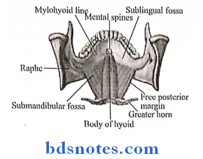
Mylohyoid Muscle Origin:
- Mylohyoid line of the mandible extending from the last molar tooth almost to the symphysis menti
Mylohyoid Muscle Insertion:
- Anterior & middle fibres- inserted into median raphe between mandible & hyoid bone
- Posterior fibres inserted into body of the hyoid bone
Read And Learn More: BDS Previous Examination Question And Answers
Mylohyoid Muscle Relations:
1. Superficial relations:
- Anterior belly of the digastric
- Superficial part of the submandibular gland
- Submental branch of the facial artery
- Mylohyoid vessels & nerve
2. Mylohyoid Muscle Deep relations:
- Hypoglossus & structures between Mylohyoid & Hypoglossus
- Styloglossus muscle
- Lingual nerve
- Submandibular ganglion
- Deep part of the submandibular gland & its duct
- Hypoglossal nerve
- Genioglossus & structures between Genioglossus & Mylohyoid
- Lingual nerve
- Submandibular duct
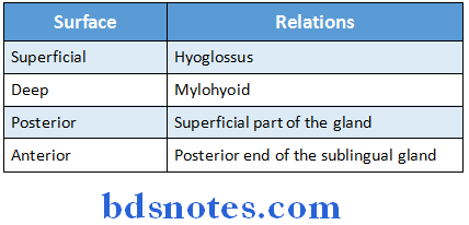
- Sublingual gland
- Third part of the lingual artery & its sublingual branch
- Hypoglossal nerve
- Geniohyoid muscle
- Structures deep to posterior free border of Mylohyoid
- Lingual nerve
- Deep part of submandibular gland
- Hypoglossal nerve
Mylohyoid Muscle Nerve Supply:
- Mylohyoid nerve, a branch of inferior alveolar nerve
Question 2. Describe the anatomy, histology, blood supply & nerve supply of Submandibular salivary gland
Answer:
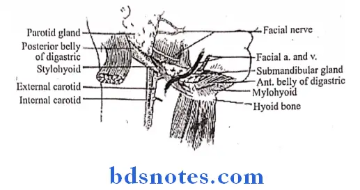
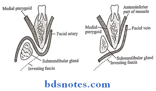
Submandibular Salivary Gland Anatomy:
- It is a large salivary gland situated in the anterior part of the digastric triangle
- It is roughly J-shaped structure being indented by the posterior border of the Mylohyoid
- This divides it into large superficial part & small deep part
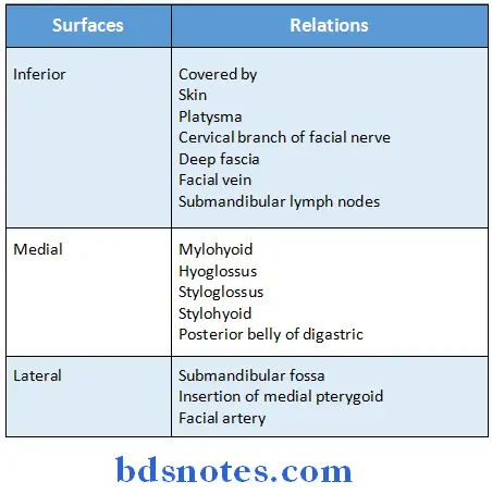
1. Submandibular Salivary Gland Superficial part:
-
-
- It fills the digastric triangle
- It has 3 surfaces
-
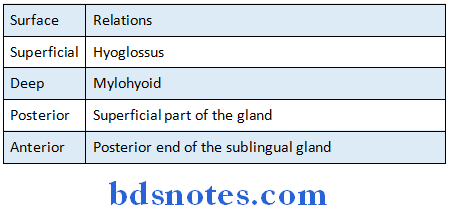
2. Submandibular Salivary Gland Deep part:
- It is related
Submandibular Salivary Gland Histology:
- Submandibular gland contains serous end peices & mucous tubules
- Serous end peices contains abundant secretory granules, spherical nucleus & basophilic cytoplasm Mucous secretory cells are filled with pale staining secretory material & little cytoplasm
- Its nucleus is compressed & contains densely stained chromatin
- The lumina of mucous tubules are larger
- The Intercalated & Striated ducts are less in number
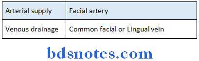
Submandibular Salivary Gland Blood supply:
Submandibular Salivary Gland Nerve supply:
- It is supplied by branches from the submandibular ganglion
- It carries
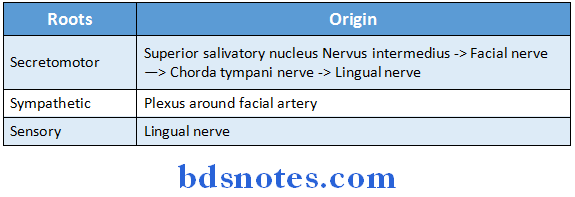
1. Secretomotor fibres:
- Its course is as follows:
- Superior salivatory nucleus → Nervus intermedius → Facial nerve → Chorda tympani nerve →→ Lingual nerve → Submandibular ganglion → Relay Post ganglionic fibres from it reaches Submandibular & Sublingual gland
2. Sensory fibres:
- It arises from the lingual nerve
3. Vasomotor sympathetic fibres:
- Arises from the plexus on the facial artery
Question 3. Give an account of extracranial course relations & branches of facial nerve. Add a note on its applied anatomy (or) Extracranial course of facial nerve (or) Facial nerve
Answer:
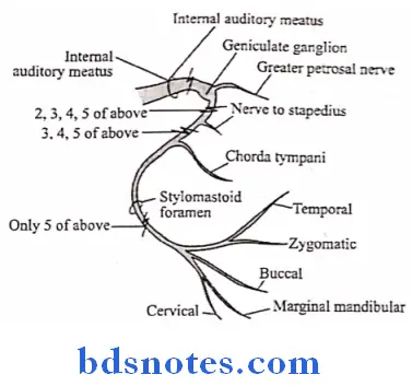
Extracranial course:
- The facial nerve leaves the skull by passing through the stylomastoid foramen
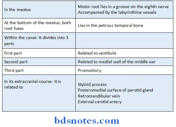
- Next in its extracranial course, it crosses the lateral side of the base of the styloid process
- Enters the posteromedial surface of the parotid gland
- Crosses the Retromandibular vein & external carotid artery
- Behind the neck of the mandible, it divides into its five terminal branches which emerge along the anterior border of the parotid gland
Extracranial Relations:
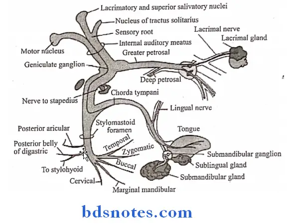
- The facial nerve is attached to the brainstem by two roots- motor & sensory
Extracranial Branches:
1. Within the facial canal
- Greater petrosal nerve
- Nerve to the stapedius
- Chorda tympani nerve
2. At its exit from stylomastoid foramen
- Posterior auricular
- Digastric
- Stylohyoid
3. Terminal branches
- Temporal
- Zygomatic
- Buccal
- Marginal mandibular
- Cervical
4. Communicating branches with adjacent cranial & spinal nerves
Extracranial Applied anatomy:
- Sudden paralysis of facial nerve at the stylomastoid foramen causes Bell’s palsy
- Lesion above the origin of chorda tympani nerve causes
- Bell’s palsy
- Loss of taste sensation from anterior two-third of tongue
- Lower motor neuron paralysis of it causes
- Paralysis of ipsilateral half of face
- Upper motor neuron paralysis
- Causes paralysis of contralateral lower quadrant of face only
Question 4. Write origin, course & branches of facial nerve. Add a note on dangerous area of the face
Answer:
Facial Nerve Origin & course:
1.Facial Nerve Originates in pons:
- The facial nerve is attached to the lateral surface of brainstem close to caudal border of the pons by two roots, sensory & motor
2. Enters internal acoustic meatus:
- At the bottom of the meatus, the two roots fuse to form a single trunk, which lies in petrous temporal bone
3. In the facial canal:
- The course is divided into three parts
-
- First part- Directed above the vestibule
- (Second part-present above the promontory
- Third part- Lies behind the promontory
-
-
- First bend is sharp called genu
- Second bend is gradual
-
4. Extracranial course:
- Exits skull via stylomastoid foramen
- Courses through parotid gland
- Divides into terminal branches which emerges along the anterior border of the parotid gland
Dangerous area of the face:
- The facial vein communicates with the cavernous sinus through its deep connections
- The facial vein is devoid of valves & rests directly on the facial muscles
- The movements of facial muscles facilitates the spread of emboli from the infected area of upper lip & lower part of the nose in retrograde direction & causes thrombosis of cavernous sinus
- Hence the upper lip & the adjoining nose lying between the angular & deep facial veins forms dangerous area of face
Question 5. Describe Submandibular Gland under following headings:
- Situation
- Surfaces and relations
- Nerve supply
- Applied anatomy
Answer:
Submandibular gland:
- It is large salivary gland
Submandibular Gland Situation:
- Present in anterior part of digastric triangle
Submandibular Gland Surfaces and relations:
- Submandibular gland is divided into two parts:
1. Superficial part
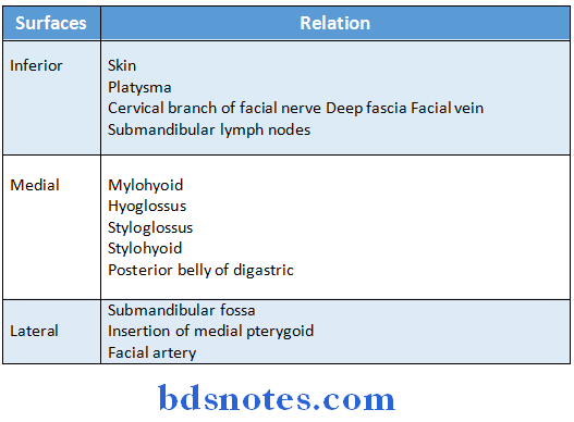
2. Deep part
Submandibular Gland Nerve supply:
- It is supplied by branches from the submandibular ganglion
- It carries
1. Secretomotor fibres
- Superior salivatory nucleus → nervus intermedius→ Facial nerve Chorda tympani nerve lingual nerve→ Submandibular ganglion
and sublingual glands
2. Sensory fibres
- It arises from the lingual nerve
3. Vasomotor sympathetic fibres
- Arises from plexus on the facial artery
Submandibular Gland Applied anatomy:
- Relay Post ganglionic fibres from it reaches submandibular
- Submandibular lymph nodes lie both within and outside the submandibular gland
- Secretion of submandibular gland is more viscous this more prone for calculi
- Excision of submandibular gland for tumour or calculus is done by incision below angle of jaw to preserve marginal mandibular branch of facial nerve
- Submandibular gland is palpated by putting one finger within the mouth and one finger outside
Question 6. Connections, course & distribution of chorda tympani nerve
Answer:
Chorda Tympani Nerve
- Chorda tympani nerve arises in the vertical part of the facial canal about 6 mm above the stylomastoid foramen
- Runs upwards & forwards in a bony canal & enters the middle ear
- Runs forwards in close relation to the tympanic membrane
- Leaves the middle ear by passing through the Petrotympanic fissure
- Passes medial to the spine of the sphenoid & enters the
infratemporal fossa - Here it joins lingual nerve
Chorda Tympani Nerve Distribution:
- Carries
- Preganglionic Secretomotor fibres to the submandibular ganglion
- This supplies Submandibular & Sublingual glands
- Taste fibres
- From the anterior two-third of the tongue
- Preganglionic Secretomotor fibres to the submandibular ganglion
Question 7. Otic Ganglion.
Answer:
- It is small, oval parasympathetic ganglion
- It is present in infratemporal fossa
Otic Ganglion Relations:
- Above- foramen ovale
- Medial- Trunk of mandibular nerve
- Lateral-Tensor veli palatini
- Front- Middle meningeal artery
- Behind- Medial pterygoid muscle
Otic Ganglion Roots:
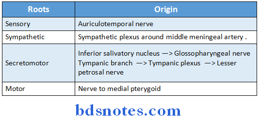
Otic Ganglion Branches:
- Secretomotor to parotid gland via Auriculotemporal nerve
- Vasomotor to parotid gland via Auriculotemporal nerve
- Motor to tensor veli palatini & tensor tympani
- Alternate route of taste pathway to anterior 2/3rd of tongue
Question 8. Submandibular Ganglion
Answer:
- It is a ganglion of parasympathetic system
- It rests on Hyoglossus muscle
Submandibular Ganglion Relations:
- Above-Lingual nerve
- Below- Deep part of Submandibular gland
Submandibular Ganglion Roots:
Submandibular Ganglion Functions:
- Supply the Sublingual, Submandibular & anterior lingual glands
Question 9. Intrapetrous part of facial nerve
Answer:
- At the bottom of the meatus, sensory & motor roots of facial nerve fuses to form a single trunk, which lies in petrous temporal bone
- In the facial canal
- The course is divided into three parts
-
- First part- Directed above the vestibule
- Second part- present above the promontory
- Third part- Lies behind the promontory
- First bend is sharp called genu
- Second bend is gradual
- Extracranial course
- Exits skull via stylomastoid foramen
Question 10. List the branches of facial nerve soon after its emergence through the stylomastoid foramen
Answer:
- Posterior auricular
- Digastric
- Stylohyoid
Question 11. Digastric muscle
Answer:
- It contains posterior & anterior bellies
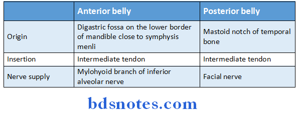
Digastric Muscle Actions:
- It depresses & retract chin in opening the mouth
- They pull hyoid bone upward & help in deglutition
Question 12. Submandibular duct
Answer:
- It is also called Wharton’s duct
- It is about 5 cm long
- Emerges at the anterior end of the deep part of the gland
- Runs forward on the hyoglossus, between the lingual & hypoglossal nerves
- In the terminal part of its course, it lies below the mucosa of the floor of the mouth
- It opens at the Sublingual caruncle or papilla just lateral to the frenulum
Question 13. Submandibular lymph nodes
Answer:
Submandibular Lymph Nodes Position:
- In the digastric triangle beneath the deep cervical fascia
Submandibular Lymph Nodes Number:
- They are three in number
-
- At anterior end of Submandibular gland
- Front of facial artery
- Behind facial artery
Submandibular Lymph Nodes Afferents:
- Center of forehead
- Medial angle of eye
- Side of nose
- Cheek & angle of mouth
- Upper lip
- Lateral part of lower lip
- Anterior two-third of tongue
- Gingiva
- Frontal, Maxillary & ethamoidal sinus
- Submental lymph node
Submandibular Lymph Nodes Efferent:
- Jugulo-omohyoid lymph nodes
- Jugulodigastric lymph nodes
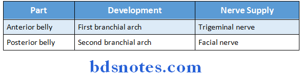
Question 14. Give the development & nerve supply of digastric muscle
Answer:

Leave a Reply