Carbohydrates General
Question 1. Define carbohydrates. Classify them and add a note on the biomedical importance of carbohydrates.
(Or)
Classification of carbohydrates
(or)
Describe the aerobic glycolysis in muscle and compute energy.
(or)
Outline the process of glycolysis. Define substrate-level phosphorylation. Give two examples.
Answer:
Definition:
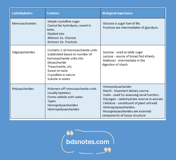
- Carbohydrates may be defined as polyhydroxy aldehydes or ketones or compounds which produce them on hydrolysis.
Read And Learn More: BDS Previous Examination Question And Answers
Classification:
- Carbohydrates are broadly classified based on the number of sugar units.
- They are:
Read And Learn More: BDS Previous Examination Question And Answers
Question 2. What are disaccharides? How will you identify them in the laboratory? What is the normal level of glucose in the blood?
Answer:
Disaccharides:
- Disaccharides are oligosaccharides containing two monosaccharide units held together by a glycosidic bond.
Properties:
- Sweet to taste
- Crystalline in nature.
- Water soluble.
Types:
1. Reducing disaccharides
- Contains free aldehyde or keto group.
- Example: Maltose, lactose.
2. Non-reducing disaccharides.
- They don’t contain any free aldehyde/keto group.
- Examples: sucrose, trehalose.
Laboratory tests for disaccharides:
1. Molisch test:
- It is a general test for the detection of carbohydrates
- The strong H2SO3 hydrolyses disaccharides to liberate monosaccharides.
- The monosaccharides get dehydrated to form furfural or hydroxyl methyl furfural which condenses with a-naphthol to form a violet-colored complex.
2. Benedict’s test:
- This is a test for the identification of reducing sugars that form enediols.
- The enediol forms of sugar reduce cupric ions of a yellow precipitate of cuprous hydroxide or a red precipitate of cuprous oxide.
3. Barfoed’s test:
- It is used to distinguish between monosaccharides and disaccharides.
- Strong reducing sugars like monosaccharide give this test positive.
4. Osazone test:
- Phenylhydrazine in acetic acid, when boiled with reducing sugars forms osazones.
- The first two carbons are involved in this reaction.
- The sugars that differ in their configuration on these carbons give the same type of osazones.
- Maltose gives sunflower-shaped osazones while lactose powder – puff shaped.
5. Sucrose hydrolysis test:
- Sucrose can be hydrolyzed by concentrated ITCl, to be converted to glucose and fructose.
- These are then reduced by Benedict’s test.
Normal blood glucose level:
- Glucose – 70 – 100 mg/dl [Normal fasting level]
Question 3. Structure and biomedical importance of disaccharides.
Answer:
Structure:
Disaccharide consists of two monosaccharide units either similar or dissimilar held together by a glycosidic bond.
Different types of disaccharides and their structures are as follows.
1. Reducing disaccharides:
- Maltose:
- It consists of two a-D-glucose units held together by a (1→4) glycosidic bond.
- It contains a free aldehyde group on C of second glucose.
- Lactose:
- It consists of P-D-galactose and P-D-glucose held together by a P(1→4) glycosidic bond.
- The anomeric carbon of C} glucose is free for reducing reactions.
2. Non-reducing disaccharides:
- Sucrose:
- It consists of a-D-glucose and p-D-fructose held by a glycosidic bond between Cl of a-glucose and C2 and P-fructose.
Question 4. Polysaccharides.
Answer:
- They are polymers of monosaccharide units with high molecular weight.
- The monosaccharides are held together by glycosidic bonds.
Properties:
- They are usually tasteless.
- They form colloids with water.
- They are linear as well as branched polymers.
Types:
1. Homopolysncchnrldes:
- These on hydrolysis yield a single type of monogenic dim ide.
- They are named according to The nature of the monosaccharide unit like.
Examples:
- Starch.
- It is a homopolymer composed of D-glucose units held by cx-glycosidic bonds.
- It consists of two polysaccharide components,
- Water soluble amylase
- Water-insoluble amylopectin.
- Dextrins.
- Inulin.
- Glycogen.
2. Hetcropolysaccharide:
- These on hydrolysis yield a mixture of a few monosaccharides.
Examples:
- Mucopolysaccharides.
- Also known as glycosaminoglycans.
- They are heleroglycans made up of repeating units of sugar derivatives, namely amino sugars and uronic acids.
- They are essential components of tissue structure
- The ground substances are principally composed of it.
- Important mucopolysaccharides include.
- Hyaluronic acid.
- Chondroitin sulfate
- Heparin.
Question 5. Name the nonreducing sugars. What are their components?(Or) Explain why sucrose is a non-reducing sugar.
Answer:
- Sucrose is a nonreducing sugar.
Components:
- Sucrose consists of a-D-glucose and p-D fructose.
- These two monosaccharides are held together by a glycosidic bond between Cl of a-glucose and C2 of p-fructose.
- The reducing groups of glucose and fructose are involved in glycosidic bonds, hence sucrose is a non-reducing sugar.
Functions:
It is the major carbohydrate produced in photosynthesis.
Employed ns sweetening agent in the food industry.
Question 6. Amylose amd amylopectin.
Answer:
1. Amylose:
- It is water soluble.
- It is a long unbranched chain with 200 – 1000 D-glucose units held by 2 -glycosidic linkages.
- Starches are hydrolyzed by amylase.
2. Amylopectin:
It is water-insoluble.
It is a branched chain with (l→6) glycosidic bonds at the branching points and a (1 → 4) linkages everywhere else.
It consists of 80-85% of total starch.
Glycogen:
- It is a tiring carbohydrate reserve in animals.
- It is present in.
- Animals
- Plants.
- Liver, muscle, brain – in humans.
Its structure consists of repeated units of glucose.
These units are joined together by a (1→4) glycosidic bond and a (1→6) glycosidic bond at branching points.
Question 7. Heteropolysaccharides- give example, composition and functions (or) Mucopolysaccharides
Answer:
- Mucopolysaccharides are heteroglycans made up of repeated units of sugar derivatives namely amino sugars and uronic acids
- They are essential components of tissue structure
- Important mucopolysaccharides include:
Question 8. Name four mucopolysaccharides in the body.
Answer:
Mucopolysaccharides:
- Hyaluronic acid
- Chondroitin 4 – sulfate
- Heparin
- Dermatan sulfate
- Keratan sulfate
Question 9. Name three glycosaminoglycans or mucopolysaccharides in the body. Mention its significance.
Answer:
Question 10. Composition and functions of two homopolysaccharides. (or) Name the cane sugar. Mention its composition.
Answer:
Question 11. What is milk sugar? Mention its composition.
Answer:
- Lactose is the milk sugar.
Composition:
- Lactose consists of p-D-galactose and p-D glucose which are held together by a p (1→ 4) glycosidic bond.
Question 12. Mutarotation.
Answer:
Definition:
- It is defined as the change in the specific optical rotation representing the interconversion of a and p forms of D-glucose to an equilibrium mixture.
Equilibrium mixture contains.
- 63% – P anomer
- 36% – a – anomer
- 1 % – open-chain form.
Question 13. Heparin. (or) Glycogen – structure and function/structure of animal starch.
Answer:
- It is one of the important mucopolysaccharides present in the body.
Composition:
It is composed of alternating units of N – sulfo – D – glucosamine 6-sulfate and glucuronate 2 – sulfate.
Functions:
- Acts as anticoagulant.
- Helps in the release of the enzyme lipoprotein lipase which clears the turbidity of Iipemic plasma.
Question 14. Detection of fructose in urine.
Answer:
Fructose is a reducing sugar and ketohexoses.
1. Test for reducing property:
- Benedict’s test:
- Fructose form enediols
- The enediol forms of sugars reduce cupric form to a yellow precipitate of cuprous hydroxide or a red precipitate of cuprous oxide.
- Barfoed’s test:
Similar to Benedict’s test but carried out in a slightly acidic medium.
2. Test for detection of fructose as ketohexoses.
- Seliwanoffs test:
- Concentrated HCI dehydrates ketohexoses to form furfural derivatives.
- This condenses with resorcinol to give a cherry red complex.
- Toulge/s test:
- The furfural derivatives of ketohexoses condense with urea in the presence of stannous chloride to give blue color.
- Rapid furfural test:
- Ketohexoses are converted to furfural derivatives by 11C1.
- This forms a purple color complex with α-naphthol.
Question 15. Epimers.
Answer: If two monosaccharides differ from each other in their configuration around a specific carbon atom, they are referred to as epimers.
Examples:
- The interconversion of epimers is known as epimerization.
Question 16. Osazone.
Answer:
- D Phenylhydrazine when boiled with reducing sugars, forms some.
Question 17. What is ketoacidosis? Name two conditions leading to it.
Answer:
- Ketoacidosis is a metabolic state associated with high concentrations of ketone bodies, formed by the breakdown of fatty acids and the deamination of amino acids.
- It is marked by extreme and uncontrolled ketosis.
Conditions leading to ketoacidosis:
- Uncontrolled diabetes mellitus
- Prolonged alcoholism
- Starvation
Question 18. Describe the nutritional importance of dietary fibers
Answer:
Importance of dietary fibers:
- Prevents constipation – helps to maintain normal motility of GIT
- Eliminates bacterial toxins
- Decreases occurrence of GIR cancers
- Improves glucose tolerance
- Reduces plasma cholesterol level
- Satiety valve – gives the sensation of stomach fullness
Question 19. What are anomers? Explain with examples
Answer:
Anomers:
- The a and (3 cyclic forms of D- glucose is known as anomers
- They differ in configuration around the C1 atom
- In an anomer, the -OH group held by anomeric carbon is on the opposite side of group CH2OH of the sugar ring, and vice versa for the p anomer
Question 20. Enumerate the difference between starch and glycogen.
Answer:
Question 21. Name 2 disaccharides and give their composition
Answer:
Disaccharides:
- Disaccharides are oligosaccharides containing two monosaccharide units held together by glycosidic bonds.
Examples:
- Maltose
- Consist of two α – D- galactose units held together by a (1→4) glycosidic bond
- Lactose
- Consist of β – D- galactose and β – D-glucose held together by β (1→4) glycosidic bond
- Sucrose
- Consists of β – D-glucose and β- D-fructose held together by a glycosidic bond between Cl of ci- glucose and C2 of β-fructose.
Question 22. Name one reducing and one non-reducing disaccharide.
Answer:
Reducing disaccharide – maltose and lactose Non-reducing disaccharide – sucrose
Question 23. What are the causes and metabolic derangements of metabolic acidosis?
Answer:
Question 24. Lactose intolerance.
Answer:
- Lactose intolerance is due to a defect in the enzyme lactase
- Continuous consumption of lactose by lactose-intolerant individuals leads to flatulence.
- This includes increased intestinal motility, cramps, and irritation.
- Its treatment includes the elimination of lactose from the diet
- Restriction of milk and dairy products helps in such cases.
Digestion And Absorption
Question 1. Discuss digestion and absorption of carbohydrates in the gastrointestinal tract.
Answer:
Digestion of carbohydrates:
1. In the mouth:
- During mastication, salivary amylase acts on starch and causes breakage of the a-glycosidic bond.
- Products formed are dextrins, maltotriose, and maltose.
2. In the stomach:
- Carbohydrate is not digested in the stomach due to the high acidity present in them.
3. In the intestine:
- The acidic constituents from the stomach are neutralized by the bicarbonate content of the pancreas.
- The pancreatic amylase acts and a-1, 4 -glycosidic bonds of starch and causes its breakage.
- The resultant products are disaccharides and oligosaccharides.
- The enzyme oligosaccharides act on oligosaccharides and disaccharidases act on disaccharides.
- This converts both these carbohydrates to monosaccharides.
- It occurs at the mucosal lining of the upper jejunum.
Absorption of carbohydrates:
- Monosaccharides produced by digestion are glucose – 80%, fructose, and lactose.
- The absorption takes place in the duodenum and upper jejunum.
1. Absorption of glucose:
- Glucose is transported by means of sodium co-transport.
- Glucose first combines with a carrier and forms glucose- a carrier complex.
- This moves the glucose across the cell membrane and releases glucose inside the cell.
- The glucose and Na+ have the same carrier called sodium-dependent glucose transporter.
- To concentrate the glucose within the cell, the carrier is coupled to a source of energy which is obtained by the binding process of sodium ion and glucose molecule to the carrier protein.
- The glucose is then transported into the interstitial space and then the blood capillaries.
2. Absorption of fructose:
- Fructose is transported by facilitated diffusion mediated by a carrier.
- Inside the cell, most of it is converted to glucose.
3. Absorption of pentoses:
- Occurs by simple diffusion.
Metabolism
Question 1. Define glycolysis, describe the reactions of its mention its significance.
Answer:
Glycolysis:
Also known as Embden – Meyerhof pathway.
It is derived from the Greek words “glycose”. sweet; “lysis” – dissolution/breakdown.
Definition:
It is defined as the sequence of reactions converting glucose/glycogen to pyruvate/lactate with the production of ATP.
It takes place in all cells of the body.
Reactions:
- The glycolysis pathway can be divided into three distinct phases.
- Energy investment phase
- Splitting phase.
- Energy generation phase.
Significance:
- Glycolysis is very essential in skeletal muscle during strenuous exercise,
- Glycolysis hi the erythrocytes leads to lactate production.
- The brain, retina, skin, renal medulla, and GIT drive most of their energy from glycolysis.
- The occurrence of glycolysis is very much elevated in rapidly growing cancer cells.
Reactions:
The glycolysis pathway can be divided into three distinct phases.
- Energy investment phase
- Splitting phase.
- Energy generation phase.
Energies:
Question 2. Describe anaerobic glycolysis. Add a note on its bioenergetics.
Answer:
Anaerobic glycolysis:
- Glycolysis occurring in the absence of oxygen is called anaerobic glycolysis.
- Lactate is the end product of it.
Pathway:
Bioenergetics:
Thus, two ATP are synthesized in anaerobic glycolysis.
Question 3. Discuss the steps of glycolysis. Indicate the sites at which ATPs are formed.
Answer:
Refer to Question No. 5 of the Long Essay.
Question 4. Describe the citric acid cycle and its energetic, (or) Give an account of the steps of the citric acid cycle. Explain why it is known as a terminal oxidative pathway. (or) Citric acid cycle. (or) Starch and glycogen
Answer:
Citric acid cycle:
Locations:
In the mitochondrial matrix, in close proximity to the electron transport chain.
Features:
- It is a cyclic process.
- It involves the oxidation of acetyl CoA to Co2 and water.
- It involves the combination of a 2-carbon acetyl CoA with a 4-carbon oxaloacetate to produce a 6-carbon tricarboxylic acid, citrate.
Reactions:
The resultant oxalacetate can combine with another molecule of acetyl CoA and continues the cycle.
Energetics:
- Each NADH produces 3 ATP through the electron transport chain. 3 NADH is produced in the TCA cycle.
- Each FADH2 produces 2 ATP through the electron transport chain. 1 FADH2 is produced in the TCA cycle.
- Substrate-level phosphorylation produces 1 ATP.
- One substrate-level phosphorylation occurs.
- Thus, the total ATP formed is.
Question 5. What are the various sources for the formation of acetyl CoA in the body and how it is oxidized to CO2 and water?
Answer:
Sources of Acetyl CoA:
- In mitochondrial matrix.
- Oxidation of pyruvate and fatty acids.
- Degradation of the carbon skeleton of certain minor acids.
- Ketone bodies.
Oxidation of Acetyl CoA:
- Acetyl CoA is oxidized to C02 and water through the citric acid cycle.
- Refer to Question No. 7 of the Long Essay.
Question 6. Describe the HMP pathway.
Answer:
Hexose monophosphate pathway:
- Pentose phosphate pathway.
Features:
- Alternative pathway to glycolysis and TCA cycle for the oxidation of glucose.
- It is more anabolic in nature.
- No ATP is directly utilized or produced.
Location:
- Located in the cytosol.
Reactions:
1. Oxidative phase: [It is concerned with the interconversion of monosaccharides.
- Initially ribulose 5 – phosphate is converted to.
- Xylulose 5 – phosphate by an epimerase
- Ribose 5 – phosphate by ribose 5- phosphate
keto isomerase.
Next, the following reactions occur and the carbon atoms are interconverted.
2. Non-oxidative Phase:
- It is concerned with the interconversion of monosaccharides.
- Initially ribulose 5 – phosphate is converted to.
- Xylulose 5 – phosphate by an epimerase
- Ribose 5 – phosphate by ribose 5- phosphate keto isomerase.
Next, the following reactions occur and the carbon atoms are interconverted.
- Fructose 6 – phosphate and glyceraldehydes 3 – phosphate can be further catabolized through glycolysis and citric acid cycle.
Significance:
- The HMP pathway generates two important products.
Question 7. Explain the glycogen synthesis in the liver and its hormonal regulation.
Answer:
Steps:
1. Synthesis of UDP glucose:
2. Primer – importance:
- Required for initiation of glycogen synthesis.
- In the absence of glycogen primer, the hydroxyl group of amino acid tyrosine of glycogen accepts glucose from UDPG.
- The enzyme glycogen initiator synthase transfers the first molecule of glucose to glycogen.
- Next glycogenin takes up a few glucose residues to form a primer.
3. Glycogen synthesis:
Enzyme involved:
- Glycogen synthase
Action:
- Transfers glucose from UDP – glucose to the non-reducing end of glycogen to form a – 1, 4 linkages.
4. Formation of branches in glycogen:
Regulation of glycogenosis:
- Glycogenesis is regulated by cAMP and by glycogen synthase.
1. Glycogen synthase enzyme.
- It exists in two forms.
- Glycogen synthase ‘a’ not phosphorylated, active form.
- Glycogen synthase ‘b’ phosphorylated, inactive, form, ‘a’ form can lie converted to ‘b’ form by phosphorylation by a cAMP-dependent protein kinase enzyme.
- This inactivates glycogen synthase and results in the inhibition of glycogenes s.
- ‘b’ form is converted to active ‘a’ form by protein phosphatase I.
- Active ‘a’ from, thus, causes glycogenesis.
1. cAMP-dependent protein kinase.
Hormones regulating cAMP:
Question 8. Describe glycogenolysis, the role of hormones in glycogenolysis, and its functional significance.
Answer:
Glycogenolysis:
- It is the degradation of stored glycogen in the liver and muscle.
- Glycogen is degraded by breaking ci -1,4 & a -1,6 glycosidic bonds
Reactions:
Role of hormones:
- Epinephrine and glucagon promote glycogenolysis by converting inactive phosphorylase ‘b’ to active phosphorylase ‘a’
- Elevated glucagon/epinephrine increases glycogen degradation
- Elevated insulin level increases glycogen synthesis
Functional significance:
- Glycogenolysis regulates glucose levels in the blood
- Plays an important role in the fight or flight response.
- In monocytes, it serves to provide an immediate source of glucose 6 – phosphate for glycolysis to provide energy for muscle contraction.
- In hepatocytes, glycogenolysis helps to provide glucose for uptake by cells.
- In it the phosphate group of glucose 6 – phosphate is removed by the enzyme glucose 6 – phosphatase and the free glucose exits the cell via GLUT 2 (Glucose Transporter – 2)
Question 9. Describe the following aspects of the citric acid cycle.
- Reactions
- Energetics
- Regulation
- Functions
Answer:
Citric acid cycle:
It is a cyclic process that involves the oxidation of acetyl CoA to carbon dioxide and water.
Reactions:
Energetics:
- Each NADH produces 3 ATP through the electron transport chain. 3 NADH is produced in the citric acid cycle
- Each FADH2 produces 2 ATP through the electron transport chain. 1 FADH2 is produced in the citric acid cycle.
- Substrate-level phosphorylation produces one ATP
Thus, the total ATP produced is:
Regulation:
- The citric acid cycle is regulated by three simple mechanisms
- Substrate availability
- Product inhibition
- Competitive feedback inhibition
- All regulatory enzymes of the citric acid cycle are inhibited by NADH
- ATP is an allosteric inhibitor of pyruvate dehydrogenase and isocitrate dehydrogenase
Functions:
- It is the most important pathway for the energy supply to the body
- It is the final oxidative pathway for carbohydrates, fats, and amino acids
- It provides many intermediates required for the synthesis of amino acids, glucose, heme, etc.
- It synthesizes
- Aspartate from oxaloacetate
- Glutamate from a-ketoglutarate
- Porphyrins and heme from succinyl CoA
- Fatty acids, steroids from acetyl CoA
Question 10. Glycerol is not a carbohydrate – justify the statement.
Answer:
Glycerol is a three-carbon alcohol
- It has the following structure.
- Each carbon is bonded to four groups and each oxygen to two groups.
- It contains multiple hydroxyl groups.
- These form attachment sites for fatty acid chains and result in triglycerides.
- However, it lacks either aldehyde or ketone groups as present in carbohydrates.
- Thus, it is not a carbohydrate, but however, the body can use it to make glucose.
Question 11. Significance of citric acid cycle.
Answer:
- It is the most important metabolic pathway for the energy supply to the body.
- About 65 – 70% of the ATP is synthesized in the Krebs cycle.
- It is the final common oxidative pathway for carbohydrates, fats, and amino acids.
- It provides many intermediates required for the synthesis of amino acids, glucose, heme, etc.
- It is the most important central pathway connecting almost all the individual metabolic pathways.
- It is both catabolic and anabolic in nature.
- It synthesizes the following.
- Separate from oxalacetate and glutamate from a-ketoglutarate are used for the synthesis of other nonessential amino acids, purine, and pyrimidines.
- Synthesis of porphyrins and heme from succinyl CoA.
- Biosynthesis of fatty acids, steroids, etc from acetyl CoA.
Question 12. Gluconeogenesis/Describe Irreversible steps of glycolysis.
Answer:
Definition:
The synthesis of glucose from non-carbohydrate compounds is known as gluconeogenesis.
Location:
- It is located in the cytosol
- Mostly occurs in the liver
Reactions:
- Closely related to the reversed pathway of glycolysis.
- Besides, it overcomes the irreversible steps of glycolysis,
- The irreversible steps of glycolysis are:
Conversion of pyruvate to phosphoenol private.
- It occurs in two steps.
- Pyruvate converted to oxaloacetate
- It occurs in mitochondrial by the enzyme pyruvate carboxylase in the presence of ATP and CO2.
- As oxaloacetate cannot diffuse out of the mitochondria, it is converted to malate by malate dehydrogenase and transported to cytosol.
- Oxaloacetate is converted to phosphoenol pyruvate.
It occurs in the mitochondrial matrix as well s cytosol by the enzyme phosphoenolpyruvate carboxykinase.
2. Conversion of fructose 1; 6 – bisphosphate to fructose 6 – phosphate.
- Fructose 1,6- bisphosphatase converts fructose 1, 6 – bisphosphate to fructose 6 – phosphate.
3. Conversion of glucose 6 – phosphate to glucose.
- It is catalyzed by glucose 6 – phosphatase.
Question 13. Glycogen Storage Disorders
Answer:
Definition:
- The metabolic defects concerned with glycogen synthesis and degradation are referred to as glycogen storage disorders.
Causes:
- Enzyme defect
- Hereditary
Types:
Question 14. Galactosemia.
Answer:
It is a rare congenital disease in infants.
Causes:
- Galactose 1 – phosphate uridyltransferase deficiency.
- Impairs galactose metabolism which leads to.
- Galactosemia – increased galactose level in blood.
- Galactosuria – increased galactose level in urine.
- Production of galactitol from galactose
- Galactitol causes.
- Cataract
- Impaired function of liver, kidney, and nervous tissue.
- Accumulation of galactose 1 -phosphate – causes.
- Depletion of inorganic phosphate from the liver.
- Impairment of liver, kidney, and nerve function.
Other symptoms:
- Loss of weight
- Hepatosplenomegaly
- Jaundice
- Mental retardation
- Amino aciduria
- Albuminuria Treatment:
- Intake of diet which lacks galactose and lactose.
2. Galactokinase deficiency – lends to
- Galactosemia.
- Development of cataracts at a very early age at birth or within a year.
Question 15. Describe glycogenolysis and glycogenesis.
Answer:
Glycogenolysis:
- It is the degradation of stored glycogen in the liver and muscle
- Glycogen is degraded by breaking a – 1,4 & a – 1,6 glycosidic bonds
Reactions:
Glycogenesis:
It is a process of glycogen synthesis
Steps:
1. Synthesis of UDP glucose
Glucose is converted to glucose 6 – phosphate by hexokinase/ glucokinase
↓
Conversion of glucose 6 – phosphate to glucose 1 – phosphate by phosphoglucomutase
↓
Synthesis of uridine diphosphate glucose (UDPG) from glucose 1 – phosphate by UDP glucose phosphorylase
1. Primer
Enzyme glycogen initiator synthase transfers the first molecule of glucose to glycogen.
↓
Glycogenin takes up a few glucose residues to form a primer.
2. Glycogen synthesis
Transfer of glucose from UDP – glucose to the reducing end of glycogen to form a – 1,4 linkages by enzyme glycogen synthase.
3. Formation of branches of glycogen
Question 16. Write reactions of glycogenolysis. What is the significance of this pathway?
Answer:
Significance:
- Glycogenolysis regulates glucose levels in the blood
- Plays an important role in the fight or flight response
- In monocytes, it serves to provide an immediate source of glucose 6 – phosphate for glycolysis to provide energy for muscle contraction.
- In hepatocytes, glycogenolysis helps to provide glucose for uptake by cells.
- In it the phosphate group of glucose 6 – phosphate is removed by the enzyme glucose 6 – phosphatase and the free glucose exits the cell via GLUT 2 (Glucose Transporter – 2)
Question 17. Reactions of glycolysis
Answer:
- The glycolysis pathway can be divided into three phases
- Energy investment phase
- Splitting phase
- Energy generation phase
Question 18. Significance of glycolysis.
Answer:
- Glycolysis is very essential in skeletal muscle during strenuous exercise.,
- In the erythrocytes, it leads to lactate production.
- The brain, retina, skin, renal medulla, and GIT derive most of their energy from glycolysis.
- It is very much elevated in rapidly growing cancer cells.
Question 19. How much ATP is produced in aerobic and anaerobic glycolysis?
Answer:
Question 20. Citric acid cycle.
Answer:
- It is a cyclic process involving the oxidation of acetyl CoA to C02 and water.
- It involves the combination of 2C acetyl CoA with a 4C oxaloacetate to produce 6C tricarboxylic acid, citrate.
Synonyms:
- Terminal oxidative pathway.
- TCA cycle.
- Kreb’s cycle.
Question 21. Write four metabolic fates of pyruvic acid.
Answer:
The Fate of pyruvic acid:
1. In aerobic condition.
- Pyruvic acid is converted to acetyl CoA in the presence of oxygen.
- This acetyl CoA enters into the TCA cycle.
2. In gluconeogenesis.
- Pyruvic acid is converted to glucose/glycogen.
- This is used in gluconeogenesis.
3. Transamination.
- Pyruvic acid is converted to alanine by transamination.
4. In anaerobic conditions.
- Pyruvic acid is converted to lactic acid during anaerobic glycolysis by lactate dehydrogenase enzyme.
Question 22. Mention two inhibitors in the glycolytic pathway. How do they act?
Answer:
Question 23. Define gluconeogenesis. Mention the key enzymes of it.
Answer:
Definition:
- The synthesis of glucose from non-carbohydrate compounds is known as gluconeogenesis.
Key enzymes:
- Pyruvate carboxylase
- Fructose 1, 6 – bisphosphate
- Glucose 6 – phosphatase.
Question 24. Significance of HMP pathway.
Answer:
- The HMP pathway generates two important products.
1. Pentoses:
Used for the synthesis of nucleic acids and nucleotides.
2. NADPH:
- Required for reductive biosynthesis of fatty acids and steroids.
- Used in the synthesis of certain amino acids.
- Brings about the detoxification of drugs.
- Helps in phagocytosis.
- Essential to preserve the integrity of the RBC membrane.
Question 25. Various glycogen storage disorders.
Answer:
The metabolic defects concerned with glycogen synthesis and degradation are referred to as glycogen storage disorders. They occur due to enzyme defects.
Question 26. Von Gierker’s disease
Answer: It is a metabolic defect concerned with the glycogen synthesis
Cause:
Glucose -6- phosphatase defect
Clinical Features:
- Enlarged liver and kidney
- Fasting hypoglycemia
- Lactic acidaemia
- Hyperlipidaemia
- Ketosis
- Gouty arthritis
Glucose Homeostasis
Question 1. Name hormones that increase the blood level with their mechanism. (or) Describe the regulation of blood glucose and add a note on disorders of blood glucose regulation.(or) Glycolysis.
Answer:
Regulation of blood glucose:
- The fasting blood glucose level in 70-100 nig/dl
- Following the ingestion of a carbohydrate meal, the blood glucose level is within normal range.
- It depends on the amount of glucose entering into circulation from processes like diet, glycogenolysis gluconeogenesis, etc., and the amount that is utilized for metabolic purposes.
- Kidneys and hormones play an important role in the regulation of blood glucose.
1. Role of the kidney:
- Glucose is filtered, reabsorbed anil returned back to circulation by the kidney.
- When the glucose concentration increases above 160 – 180 mg/dl, it is exerted in urine.
2. Role of hormones:
Disorders:
1. Hyperglycemia:
- It occurs when the blood glucose concentration is less than 45 mg/ill
- Clinical features:
- Headache, seizures, anxiety, coma.
- Sweating, slurred speech.
Types:
Observed in an individual with elevated insulin secretion following a meal
2. Diabetes mellitus:
- It is a metabolic disease characterized by hyperglycemia.
Types:
- Insulin-dependent diabetes mellitus (Type I)
- Mainly occurs in children with so-called juvenile-onset diabetes.
- Characterized by total deficiency of insulin due to the destruction of p-cells of the pancreas.
- Noninsulin-dependent diabetes mellitus-type II.
- Occurs in adults.
- This occurs due to genetic and environmental factors.
Causes:
-
- Obesity
- Decrease in insulin receptors.
3. Glycosuria:
One of the symptoms of diabetes mellitus.
Types:
- Renal glycosuria.
- It is a benign condition due to a reduced renal threshold for glucose.
- Alimentary glycosuria.
-
- In certain individuals, blood glucose level rises rapidly after meals.
- This glucose is excreted in urine and this condition is called alimentary glycosuria.
Question 2. Oral glucose tolerance test.
Answer:
Definition:
The diagnosis of diabetes made on the basis of individuals’ response to the oral glucose load is called the glucose tolerance test (GTT).
Preparation of the subject:
- The subject is provided with a carbohydrate-rich diet for at least 3 days prior to the test.
- Drugs affecting carbohydrate metabolism are discontinued for at least 2 days.
- Strenuous exercise and smoking should be avoided on the previous day of the test.
- The subject should be in an overnight fasting state.
Procedure:
- Conducted preferably in the morning.
- Fasting blood and urine sample is taken.
- Next, the subject is given 75 g of glucose dissolved in about 300 ml of water, given orally.
- Blood and urine samples are collected at the interval of 30 minutes for 2 hours.
- The values obtained are plotted on a graph.
Interpretation:
Question 3. Glycosuria.
Answer:
- It is the condition of glucose excretion in urine.
- Glucose appears in urine when the plasma glucose concentration exceeds the renal threshold for glucose.
Types:
1. Renal glycosuria.
- It is a benign condition.
- Occurs due to a reduced renal threshold for glucose.
- It is unrelated to diabetes.
2. Alimentary glycosuria.
- In certain individuals, blood glucose rapidly increases after meals which get excreted in the urine.
- This is known as alimentary glycosuria.
- It is observed in.
- Normal individuals.
- Individuals with.
- Hepatic diseases
- Hyperthyroidism
- Peptic ulcer.
Question 4. Normal GTT curve.
Answer:
Procedure:
Curve:
- Glucose concentration in the fasting state is the norm.il individuals obtained Is. 75 – 110 mg/dl.
- On oral glucose administration, the concentration increases rapidly and reaches the peak value of HO mg/dl.
- This value is reached within one hour
- In the next hour, it decreases and comes back to normal
- His curve is obtained from tire blood samples.
Question 5. Metabolic changes in diabetes mellitus
Answer:
Diabetes mellitus is associated with several metabolic alterations.
The most important of them are:
Glucose uptake
Glucose production
Major Metabolic Changes In Diabetes Mellitus:
Absolute or relative insulin deficiency
↓
Multiple metabolic effects
Curve:
- Glucose concentration in fasting state in normal individuals obtained is 75 – 110 mg/ dl.
- On oral glucose administration, the concentration increases rapidly and reaches a peak value of 140 mg/dl.
- This value is reached within one hour.
- In the next hour, it decreases and comes back to normal.
- This curve is obtained from the blood samples.
Question 5. Metabolic changes in diabetes mellitus
Answer:
- Diabetes mellitus is associated with several metabolic alterations
- The most important of them are:
↓Glucose uptake
↑Glucose production
Major Metabolic Changes In Diabetes Mellitus:
Absolute or relative insulin deficiency
↓
Multiple metabolic effects
↓
Question 1. GTT.
Answer:
- It means a glucose tolerance test.
- The diagnosis of diabetes is made on the basis, of individuals’ response to the oral glucose load n n-her-d as an oral glucose tolerance test.
Value:
1. In normal individuals.
- Fasting glucose concentration – 75 – 110 mg/dl.
2. In patients with impaired glucose tolerance.
- Fasting glucose – 110 – 26 mg/dl.
- Postpardinal glucose – 140 – 200 mg/dl.
3. In diabetic patients.
- Fasting glucose – exceeds 126 mg/dl.
- Postpardinal glucose – exceeds 200 mg/dl.
Question 3. What is glycosuria? Mention the renal threshold for glucose.(or) Compare the GTT curve with different diabetic conditions (or) Renal glycosuria.
Answer:
Glycosuria:
- The excretion of glucose in the urine is called glycosuria.
The renal threshold for glucose:
- If the level of glucose in the blood is above 160 – 180 mg/dl it is excreted in urine.
- This value is referred to as the renal threshold for glucose.
Question 5. Mention the functions of insulin and glucagon.
Answer:
Question 6. Name the hormones that increase blood glucose.
Answer:
Hormones that increase blood glucose are:
- Glucagon
- Epinephrine
- Thyroxine
- Glucocorticoids
- Growth hormone
- 6ACTH.
Leave a Reply