Orthodontic Study Models – Study Casts
Study casts are essential diagnostic aids.
- Orthodontic study models are accurate plaster reproduction of the teeth and their surrounding soft tissues.
- Study models are three-dimensional view of the maxillary and mandibular dental arches.

“Understanding the role of orthodontic study models in treatment planning: Q&A explained”
Dental Study Models
Read And Learn More: Orthodontics Question And Answers
Study Models In Dentistry – Use of Study Models
- They enable the study of occlusion from all aspect.
- They enable accurate measurement to be made in a dental arch. They help in measurement of arch length, arch width and tooth size.
- They help in assessment of treatment progress by dentist as well as the patient.
- They help in assessing the nature and severity of malocclusion.
- They are helpful in motivation of the patient and to explain the treatment plan as well as progress to the patient and parents.
- Study models make possible to stimulate treatment procedure on the cast such as mock surgery.
- Study models are useful to transfer records in case the patient is to be treated by another clinician.
“Importance of studying orthodontic study models for better outcomes: Questions explained”
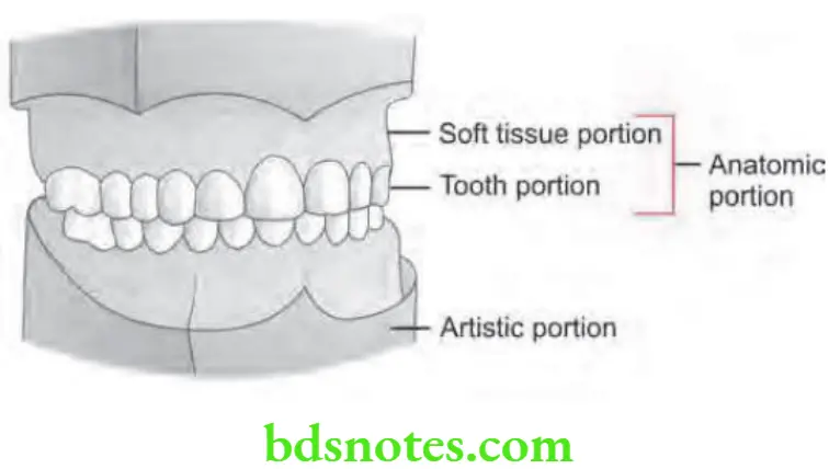
Study Models In Dentistry – Parts of a Study Model
- Anatomic portion
- Artistic portion
- Anatomic portion: The anatomic portion is that part of the study model which is the actual impression of the dental arch and its surrounding structures. Anatomic potion is usually made of stone plaster.
- Artistic portion: This part consists of a plaster base that supports the anatomic portion.
In a well trimmed study casts, ratio between anatomic and artistic portion should be 2:1. Tooth portion, sof tissue portion and artistic portion should be 1:1:1. Completed model is 13 mm in height in both anterior and posterior region.
“Differential applications of manual vs digital orthodontic study models: Questions answered”
“Common challenges in using orthodontic study models effectively: FAQs provided”
Dental Cast Models – Fabrication of Study Cast
Impression Making
- Obtain a good alginate impression for proper fabrication of orthodontic casts.
- Orthodontic study models reproduce as much of supporting structures as possible, so it is recommended to use high flnge orthodontic trays that extend deep in buccal and lingual sulci.
- Selected tray should cover last erupted molar and have clearance of 3 mm between the teeth and tray.
- A good impression shows a peripheral roll and records the muscle attchments. Retromolar pad in the mandible and tuberosity in maxilla should be included.
Disinfection of Impression
- Impression should be rinsed in water and disinfected by biocide solution to remove microorganisms, plaque, mucin and other debris which reduces the quality of surface reproduction.
- As disinfection is completed, once again impression is rinsed in water to clear residual disinfectant.
“Asymptomatic vs symptomatic effects of ignoring study model findings: Q&A”
Casting of Impression
- Rinse the impression and excess water is shaken out.
- Good quality of stone model plaster is used to pour the impression.
- It is always best to use mechanical spatulator or vacuum mixer.
“Factors influencing the accuracy of orthodontic study models: Q&A”
Basing and Trimming of the Cast
- Rubber base formers are used to pour artistic portion or the base. They confie the plaster and are fabricated to shape of the base in artistically pleasing contour.
- Tray orientation should be done in the manner that anatomic portion is in the center of rubber mould with occlusal plane parallel with the cast base of base former.
- Guidelines for trimming the cast:
- Upper model should be cut by back edge at right angles to middle line and the front surfaces are cut so that point of intersection of front surface is in line with middle line of palate.
- Sides of the model should be cut symmetrically about the middle line
- Upper model act as a guide in trimming the lower model
- By set square, back corners of upper and lower model are trimmed. Front of lower model is trimmed to the smooth curve.
- Distal corners should be cut symmetrically to middle line conveniently with models in occlusion. Sides of model are cut symmetrically about the middle line.
- Occlusal plane should be parallel to top and bottm of study casts.
- After trimming the study casts should be symmetrical. Upper study cast should have seven sides and lower study casts have six sides when viewed from occlusal plane.
“Steps to explain different types of orthodontic study models: Digital vs plaster: Q&A guide”
Finishing and Polishing
- Final fiishing of the artistic portion of dental cast is done by fie-grained waterproof sand paper.
- Removes the bubbles appear at gingival margin by small universal sealer and those in mucobuccal fold area are removed by Kingsley type scraper.
- Final polishing is done by placing the cast in soap solution for one hour and later removed and rinsed under the warm water.
- Dry the cast and buf them so that they acquire smooth and shiny surface.
- Use model storage boxes to store fiished study models for future reference.
“Early warning signs of issues detected through orthodontic study models: Common questions”
Fabrication of Study Cast Advantages
- Allows more objective assessment of malocclusion as compared to photograph and clinical examination.
- By help of study cast occlusion from lingual aspect can be seen easily.
- They are the permanent records of patient.
- Less expensive.
- Easily duplicated.
“Role of digital impressions in orthodontic study models: Questions answered”
Fabrication of Study Cast Disadvantages
- Occupies storage space.
- Can break on fall.
- Information of soft tissues should not be obtained.
- Teeth and facial profile should not be elicited.
Leave a Reply