Understanding Extradural Haematoma: Symptoms, Diagnosis, and Innovative Treatment Options
Describe features to extradural hematoma.
Answer. Following are the features to extradural hematoma
- Patient soon regain consciousness and again after 6 to 12-hour start deteriorating (Lucid interval).
- Later the patient presents with confusion, irritability, drowsiness, hemiparesis on same side of the injury.
Initially, pupillary constriction and later pupillary dilatation occurs on the same side, finally becomes totally unconscious—Hutchinson Pupils - Death can occur if immediate surgical intervention is not done.
- Features of raised intracranial pressure such as high blood pressure, bradycardia, vomiting is also seen.
Extradural Haematoma

“Extradural Haematoma After Car Accidents”
- Occasionally, convulsions may be present.
Wound and hematoma in the temporal region of scalp may be seen. - Glasgow coma scale gives clear idea about neuronal injury.
- Autonomic disturbances with bradycardia, systolic hypertension, deep and slow respiration, Cheyne Stokes ventilation.
- Cushing’s triad of raised intracranial pressure is obvious, i.e. bradycardia, hypertension, and respiratory irregularity.
- Features such as restlessness, irritability, headache, vomiting, and progressive deterioration are common.
“Risk Factors For Developing Extradural Haematoma“
Read And Learn More: Neurological and Facial Disorders: Causes, Diagnosis, and Treatment Strategies
Extradural hematoma is a serious condition that occurs when blood collects between the outer membrane of the brain and the skull. This can happen due to head injuries or other factors. Recognizing the symptoms early and understanding the diagnostic methods are crucial for effective treatment. In this article, we will break down what extradural hematoma is, how to identify it, the latest diagnostic techniques, and explore innovative treatment options available today.
Key Takeaways
- Extradural hematoma is a collection of blood outside the brain, often due to trauma.
- Symptoms can range from mild headaches to severe neurological issues, depending on the hematoma’s size and location.
- Imaging tests like CT scans are vital for diagnosing extradural hematoma quickly.
- Surgical options are available and can be minimally invasive, improving recovery times.
- Research is ongoing, focusing on new treatments and technologies for better management of extradural hematoma.
Epidural Hematoma
Defining Extradural Haematoma
What Is Extradural Haematoma?
Extradural haematoma, also known as epidural hematoma, is a serious condition that happens when blood collects between the dura mater (the tough outer membrane covering the brain and spinal cord) and the skull. This bleeding usually occurs because of a head injury, like a skull fracture. It’s a problem because the accumulating blood can put pressure on the brain, which can lead to brain damage. It’s important to get medical help right away if someone is suspected of having an extradural haematoma.
“Headache And Vomiting In Extradural Haematoma“
Causes of Extradural Haematoma
Most of the time, extradural haematomas are caused by traumatic head injuries. Here’s a breakdown of common causes:
- Skull Fractures: These are the most frequent cause, as they can tear the arteries or veins in the area.
- Arterial Bleeding: Bleeding from an artery, especially the middle meningeal artery, is a common cause. This type of bleeding can cause the haematoma to expand quickly.
- Venous Bleeding: Sometimes, the bleeding comes from veins. Venous bleeding tends to be slower than arterial bleeding.
- Other Injuries: In rare cases, an extradural haematoma can happen without a skull fracture, but this is less common.
It’s worth noting that while trauma is the main cause, certain factors like bleeding disorders or the use of blood-thinning medications can increase the risk of developing an extradural haematoma after a head injury.
Understanding Extradural Haematoma
Types of Extradural Haematoma
Extradural haematomas can be classified based on a few different factors. Understanding these types can help doctors determine the best course of treatment. Here are some ways they’re categorized:
- Location: Haematomas can occur in different areas of the skull, such as the temporal, frontal, or parietal regions. The location can affect the symptoms a person experiences.
- Size: The size of the haematoma is important. Larger haematomas can cause more pressure on the brain and may require surgery. Doctors use imaging scans to measure the size.
- Speed of Bleeding: Some haematomas expand quickly (acute), while others develop more slowly (chronic). Acute haematomas often need immediate treatment. Recognizing the need for neuraxial anesthesia is important in these situations.
“Red Flags Of Severe Extradural Haematoma”
Here’s a simple table to illustrate the different classifications:

Recognizing Symptoms of Extradural Haematoma
Acute Extradural Haematoma
Common Symptoms
Recognizing the signs of an extradural haematoma (EDH) early can really change things. The symptoms can show up pretty quickly after a head injury, sometimes within minutes or hours. It’s not always a clear-cut thing, though. Some people might have what’s called a lucid interval, where they seem okay for a bit before things take a turn.
Here’s what to watch out for:
- Headache: Often severe and persistent.
- Vomiting: Especially if it’s repetitive.
- Drowsiness: Feeling unusually sleepy or hard to wake up.
- Confusion: Seeming disoriented or having trouble thinking clearly.
It’s easy to brush off a headache or some confusion after a bump to the head, but with EDH, time is of the essence. If you notice these symptoms, especially if they’re getting worse, it’s important to get checked out right away.
Severe Symptoms
If an EDH isn’t caught early, the pressure on the brain can increase, leading to some serious problems. These symptoms are a sign that things are getting critical, and immediate medical attention is a must.
- Seizures: Uncontrolled shaking or jerking movements.
- Weakness or Numbness: Usually on one side of the body.
- Speech Problems: Difficulty speaking or understanding what others are saying.
- Loss of Consciousness: Becoming unresponsive.
- Pupil Changes: One pupil becoming larger than the other.
Traumatic Epidural Hematoma
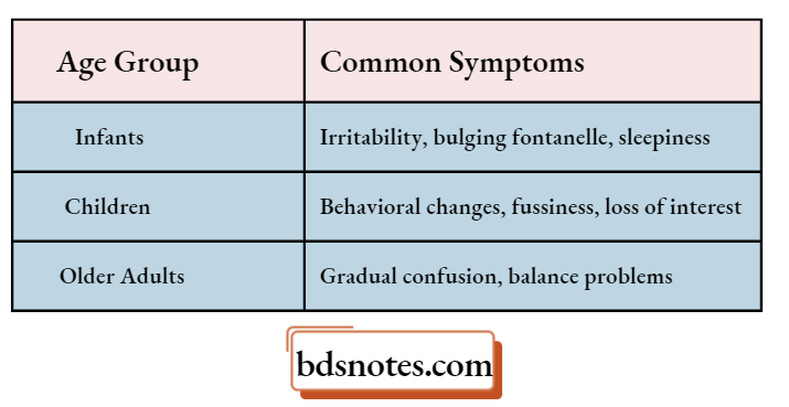
Symptoms in Different Age Groups
The way an EDH shows up can be different depending on age, especially in kids and older adults. It’s important to keep these differences in mind because what might seem like normal behavior could actually be a sign of something serious.
- Infants: Might show irritability, bulging fontanelle (soft spot on the head), or excessive sleepiness.
- Children: Could have changes in behavior, like increased fussiness or loss of interest in activities.
- Older Adults: Symptoms might be subtle, like gradual confusion or balance problems, which can be mistaken for other age-related issues.
“Early Warning Signs Of Brain Compression”
Diagnostic Approaches for Extradural Haematoma
Imaging Techniques
When it comes to figuring out if someone has an extradural haematoma, imaging is really important. It’s how doctors can actually see what’s going on inside the skull.
- CT Scans: These are usually the first thing doctors go for. They’re quick and can show a haematoma as a bright area. Sometimes, though, if the blood is the same density as the brain, it can be tricky to spot.
- MRI Scans: These give a more detailed picture than CT scans. They’re especially good at finding smaller haematomas or ones that are in tricky spots. The downside is they take longer and aren’t always available right away.
- X-Rays: While not used to diagnose extradural hematoma directly, skull X-rays can help identify fractures that may have caused the bleed.
It’s worth noting that sometimes, a CT scan might miss a small haematoma, especially early on. If symptoms are there but the CT is clear, doctors might need to do an MRI to be sure.
Extradural Haematoma Clinical Assessment
It’s not all about the machines, though. Doctors also rely on good old-fashioned clinical assessment. This means checking things like:
- Level of Consciousness: How awake and alert the person is. This is often measured using the Glasgow Coma Scale (GCS).
- Pupil Size and Reaction: Unequal pupils or pupils that don’t react to light can be a sign of pressure on the brain.
- Motor Function: Checking strength and movement in the arms and legs. Weakness on one side of the body can point to a problem on the opposite side of the brain.
A thorough neurological exam is key. It helps doctors understand the severity of the injury and where in the brain the problem might be.
Extradural Haematoma Differential Diagnosis
Extradural haematomas aren’t the only things that can cause these symptoms. Doctors need to rule out other possibilities, such as:
- Subdural Haematoma: Bleeding between the brain and its outer covering.
- Intracerebral Haemorrhage: Bleeding inside the brain tissue itself.
- Skull Fractures: Sometimes, the fracture itself can cause symptoms, even without a large haematoma.
To make sure they’re treating the right thing, doctors use imaging and clinical assessment to differentiate between these conditions. Sometimes S100B protein sampling is suggested as a screening method to identify patients at low risk for post-traumatic intracranial hemorrhage.
Traumatic Epidural Hematoma
Innovative Treatment Options For Extradural Haematoma
Extradural haematomas, while serious, have seen significant improvements in treatment approaches. It’s not just about drilling a hole anymore; doctors have a range of options, and they’re always getting better. The goal is always to minimize brain damage and get people back to their lives as quickly as possible. Let’s explore some of the current methods.
Surgical Interventions
Surgery is often the first line of defense, especially when the haematoma is large or causing significant pressure on the brain. The primary goal is to relieve that pressure quickly. The type of surgery depends on the size and location of the bleed.
- Craniotomy: This involves creating a bone flap in the skull to directly access and remove the haematoma. It’s pretty invasive, but it gives the surgeon a good view and plenty of room to work.
- Burr Hole Trepanation: For smaller, more accessible haematomas, surgeons might opt for burr holes. These are small holes drilled into the skull to drain the blood. It’s less invasive than a craniotomy, but it might not be suitable for larger clots.
- Decompressive Craniectomy: In severe cases with significant swelling, a portion of the skull might be removed temporarily to allow the brain to swell without being compressed. The bone flap is then replaced later. This is a big step, but it can be life-saving.
Surgical timing is super important. The sooner the pressure is relieved, the better the chances of a good outcome. Doctors use imaging and neurological exams to figure out the best course of action and how quickly they need to act.
“Pathophysiology Of Extradural Haematoma Explained”
Minimally Invasive Techniques
Minimally invasive techniques are becoming more popular as technology advances. They offer the potential for faster recovery and fewer complications. These methods aim to achieve the same goals as traditional surgery but with smaller incisions and less disruption to the surrounding tissues. One example is a lumbar epidural patch for related conditions.
- Endoscopic Surgery: Using a small camera and specialized instruments inserted through small incisions, surgeons can visualize and remove the haematoma. This approach is particularly useful for certain locations and smaller bleeds.
- Stereotactic Aspiration: This involves using imaging guidance to precisely target the haematoma and drain it with a needle or catheter. It’s a good option for deep-seated or difficult-to-reach clots.
- Catheter-Based Thrombolysis: In some cases, medications can be delivered directly to the clot through a catheter to dissolve it. This is less common for extradural haematomas but might be considered in specific situations.
Extradural Haematoma Pharmacological Treatments
While surgery is often necessary, medications can play a supportive role in managing extradural haematomas. These aren’t usually the primary treatment, but they can help control swelling, prevent seizures, and manage pain. Here’s a quick rundown:
- Anticonvulsants: To prevent or control seizures, which can worsen brain injury.
- Corticosteroids: To reduce swelling around the haematoma, although their use is somewhat controversial.
- Pain Management: Medications to alleviate headache and other pain symptoms.
Post-Treatment Care for Extradural Haematoma
Rehabilitation Strategies
After dealing with an extradural haematoma, getting back to normal takes time and effort. It’s not a sprint, it’s a marathon. Rehabilitation is key to regaining lost functions and improving your overall quality of life. This often involves a team of specialists, including physical therapists, occupational therapists, and speech therapists, depending on the specific deficits you’re experiencing.
- Physical therapy focuses on improving motor skills, strength, and coordination. They might have you doing exercises to help you walk, balance, and move your limbs more easily.
- Occupational therapy helps you adapt to everyday tasks. This could mean learning new ways to dress, cook, or use a computer.
- Speech therapy is important if the haematoma affected your speech, language, or swallowing abilities.
“Emerging Treatments For Extradural Haematoma”
The goal is to help you regain as much independence as possible. It’s a process, and it’s okay to have good days and bad days. The important thing is to keep working at it and celebrate small victories along the way. Remember to follow your therapist’s instructions carefully and don’t push yourself too hard, especially at first.
Monitoring For Complications
Even after successful treatment, it’s important to keep a close eye out for any potential problems. Complications can sometimes arise, even weeks or months later. Regular check-ups with your doctor are a must. They’ll likely want to do neurological exams and possibly repeat imaging scans to make sure everything is stable.
Here are some things to watch out for:
- Headaches that are getting worse or not responding to medication
- Changes in vision, speech, or coordination
- Seizures
- Weakness or numbness in your arms or legs
If you notice any of these symptoms, don’t wait. Contact your doctor right away. Early detection and treatment can make a big difference in preventing long-term problems. It’s also a good idea to keep a journal of your symptoms and any changes you notice, as this can be helpful for your doctor to track your progress and identify any potential issues. It’s important to monitor for potential problems.
Long-Term Outcomes
The long-term effects of an extradural haematoma can vary quite a bit, depending on the severity of the initial injury, how quickly you received treatment, and your overall health. Some people make a full recovery and are able to return to their normal lives without any lasting problems. Others may experience some long-term challenges, such as:
- Cognitive difficulties (problems with memory, attention, or problem-solving)
- Motor deficits (weakness, paralysis, or difficulty with coordination)
- Emotional or behavioral changes (depression, anxiety, or irritability)
It’s important to be patient with yourself and to seek support if you’re struggling. There are many resources available to help you cope with these challenges, including support groups, counseling, and rehabilitation programs. Remember that recovery is a journey, not a destination, and it’s okay to ask for help along the way. With the right support and a positive attitude, you can live a full and meaningful life even after experiencing an extradural haematoma.
Research and Future Directions in Extradural Haematoma Management
Current Studies
Right now, a lot of research is focused on better understanding how extradural haematomas (EDH) develop and affect patients. Researchers are digging into the genetic factors that might make some people more likely to experience an EDH after a head injury. There’s also a big push to refine imaging techniques so we can spot these bleeds earlier and more accurately. For example, some studies are exploring the use of advanced MRI sequences to differentiate between different types of blood clots, which could help doctors decide on the best course of action. research trends are being identified to better understand EDH.
- Investigating genetic predispositions to EDH.
- Improving the sensitivity of CT scans for early detection.
- Developing biomarkers for predicting patient outcomes.
“Case Studies On Extradural Haematoma Outcomes”
One area of intense interest is the development of non-invasive methods for monitoring patients at risk of EDH. This could involve wearable sensors that track intracranial pressure or new blood tests that can detect early signs of bleeding. The goal is to move away from relying solely on clinical symptoms, which can sometimes be misleading, and towards a more proactive approach to management.
Emerging Technologies
New technologies are changing how we deal with extradural haematomas. Artificial intelligence is starting to play a role, with algorithms being developed to analyze brain scans and help doctors make quicker, more informed decisions. Telemedicine is also becoming more important, especially in rural areas where access to specialists might be limited. This allows experts to remotely assess patients and guide local healthcare providers.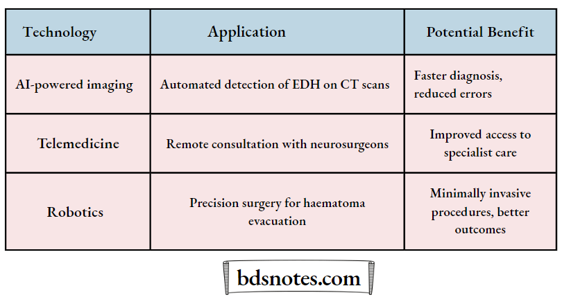
Potential Breakthroughs
Looking ahead, there are some really exciting possibilities on the horizon for treating extradural haematomas. One area is the development of new drugs that can help to stop bleeding and promote healing in the brain. Another is the use of minimally invasive surgical techniques, like endoscopic surgery, to remove blood clots with less damage to surrounding tissues. And there’s even talk of using stem cell therapy to repair damaged brain cells after an EDH.
- Development of targeted drug therapies to reduce bleeding.
- Refinement of minimally invasive surgical approaches.
- Exploration of stem cell therapy for brain repair.
“Complications Of Untreated Extradural Haematoma”
Final Thoughts on Extradural Hematoma
In summary, extradural hematoma is a serious condition that can lead to significant health issues if not addressed quickly. Recognizing the symptoms early is key, as it can make a big difference in treatment outcomes. Diagnosis often involves imaging tests like CT or MRI, which help doctors see what’s going on inside. Thankfully, there are new and innovative treatment options available that can help manage this condition more effectively. Whether through surgical intervention or other methods, the goal is to relieve pressure and restore function. If you or someone you know experiences symptoms, don’t hesitate to seek medical help right away. Awareness and prompt action can save lives.
Frequently Asked Questions
What Is An Extradural Hematoma?
An extradural hematoma is a type of bleeding that happens between the outer layer of the brain and the skull. It usually occurs after a head injury.
What Causes An Extradural Hematoma?
Extradural hematomas are often caused by head traumas, like falls or car accidents, which can damage blood vessels in the skull.
What Are The Common Symptoms Of An Extradural Hematoma?
Common symptoms include headaches, confusion, dizziness, and sometimes loss of consciousness.
“Most Common Causes Of Extradural Haematoma”
How Is An Extradural Hematoma Diagnosed?
Doctors usually use imaging tests like CT scans or MRIs to diagnose an extradural hematoma.
What Treatments Are Available For Extradural Hematoma?
Treatment often involves surgery to remove the blood and relieve pressure on the brain. In some cases, doctors might use less invasive methods.
What Should I Expect After Treatment For An Extradural Hematoma?
After treatment, patients may need rehabilitation to recover fully. Monitoring for complications is also important.
Leave a Reply