Types Of Ulcers: Symptoms And Treatment
Write short note on ulcer.
Answer. An ulcer is the break in the continuity of the covering epithelium either skin or mucus membrane due to molecular death.
Classification of Ulcer
Classification I (Clinical) of Ulcer
- Spreading ulcer
- Healing ulcer
- Non-healing ulcer
- Callous ulcer
Classification II (Based on duration) of Ulcer
- Acute ulcer: Duration less than 2 weeks
- Chronic ulcer: Duration more than 2 weeks
“What Are The Types Of Ulcers”
Classification III (Pathological) of Ulcer
Specific ulcers:
- Tuberculous ulcer
- Syphilitic ulcer
- Actinomycosis
- Meleney’s ulcer
Malignant ulcers:
- Carcinomatous ulcer
- Rodent ulcer
- Melanotic ulcer
“Symptoms Of Different Types Of Ulcers”
Non-specific ulcers:
- Traumatic ulcers
- Arterial ulcer
- Venous ulcer
- Trophic ulcer
- Infective ulcer
- Tropical ulcer
- Ulcers due to chilblain and frostbite
- Martorell’s hypertensive ulcer
- Bazin’s ulcer
- Diabetic ulcer
- Ulcers due to leukemia, polycythemia, jaundice,
collagen diseases, lymphoedema - Cortisol ulcers
“Different Types Of Ulcers Explained”
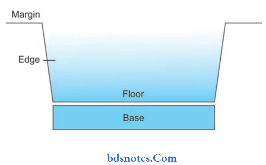
Parts of an ulcer
Margin: It may be regular or irregular. It may be rounded or oval.
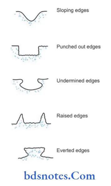
Edge: Edge is the one that connects floor of the ulcer to the margin. Different edges are:
- Sloping edge: It is seen in a healing ulcer.
Its inner part is red because of red, healthy granulation tissue. Its outer part is white due to scar/firous tissue. Its middle part is blue due to epithelial proliferation
“Best Treatments For Different Ulcer Types”
- Undermined edge is seen in a tuberculous ulcer. Disease process advances in deeper plane (in subcutaneous tissue) whereas (skin) epidermis proliferates inwards.
- Punched out edge is seen in a gummatous (syphilitic) ulcer and trophic ulcer. It is due to endarteritis.
- Raised and beaded edge (pearly white) is seen in a rodent ulcer (basal cell carcinoma). Beads are due to proliferating active cells.
- Everted edge (rolledoutedge): Itis seenina carcinomatous ulcer due to spill of the proliferating malignant tissues over the normal skin.
- Floor: lt is the one which is seen. Floor may contain discharge, granulation tissue or slough.
- Base: Base is the one on which ulcer rests. It may be bone or soft tissue.
Induration of ulcer: It is the clinical palpatory sign which means a specific type of hardness in a diseased tissue. It is seen in carcinomatous ulcers.
“Comprehensive Overview Of Ulcer Symptoms”
Investigations For Ulcer
- Study of discharge: Culture and sensitivity, acid fast bacilli study and cytology.
- Edge biopsy: Biopsy is taken from the edge because edge contains multiplying cells. Usually, two biopsies are taken.
Biopsy taken from the centre may be inadequate because of central necrosis. - X-ray of the part to look for periostitis/osteomyelitis.
- FNAC of the lymph node.
- Chest X-ray, Mantoux test in suspected case of tuberculous ulcer.
- Hemoglobin, ESR, total WBC count, serum protein estimation.
“The Role Of Imaging Tests In Diagnosing Ulcers”
Management of an ulcer
- Cause should be found and treated
- Correct the defiiencies like anemia, protein and vitamins deficiencies
- Transfuse blood, if required
- Control the pain and infection
- Investigate properly
- Control the infection and give rest to the part
- Care of the ulcer by debridement, ulcer cleaning and dressing is done daily or twice daily.
- Remove the exuberant granulation tissue
- Topical antibiotics for infected ulcers only like, silver sulphadiazine, mupirocin.
- Antibiotics are not required once healthy granulation tissues, if are formed
- Once granulates, defect is closed with secondary suturingskin graft, flaps
Leave a Reply