Tonsil
Describe tonsil under the following headings:
- Tonsil Location,
- Tonsil External features,
- Tonsil Tonsillar bed,
- Tonsil Nerve supply,
- Tonsil Arterial supply,
- Tonsil Venous drainage and
- Tonsil Applied anatomy.
“Importance Of Tonsils In Preventing Throat Infections”
Pharynx Anatomy
Answer.
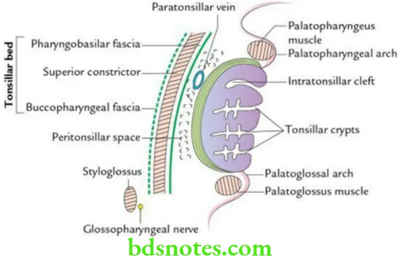
Tonsil Location The palatine tonsil is an almond-shaped mass of lymphoid tissue (dimension of about 2 cm) located in the tonsillar fossa on each side in the lateral wall of the oropharynx.
The tonsillar fossa is a triangular recess that is bound in front by the palatoglossal fold and behind by the palatopharyngeal fold.
External features of tonsil The tonsils present the following features:
- Two surfaces: medial and lateral
- Two borders: anterior and posterior
- Two ends: upper and lower
“Early Signs Of Issues With Tonsils And Throat Health”
“Risk Factors For Tonsil-Related Disorders Like Tonsillitis”
The lateral surface is covered by a sheath of condensed connective tissue called hemicapsule of the tonsil.
Tonsillar bed From deep to superficial, it is formed by:
- Pharyngobasilar fascia
- Superior constrictor muscle supplemented by palatopharyngeus
- Buccopharyngeal fascia
Tonsillar Nerve supply
- Glossopharyngeal nerve
- Lesser palatine nerves
“Understanding The Role Of Tonsils In Immune Defense”
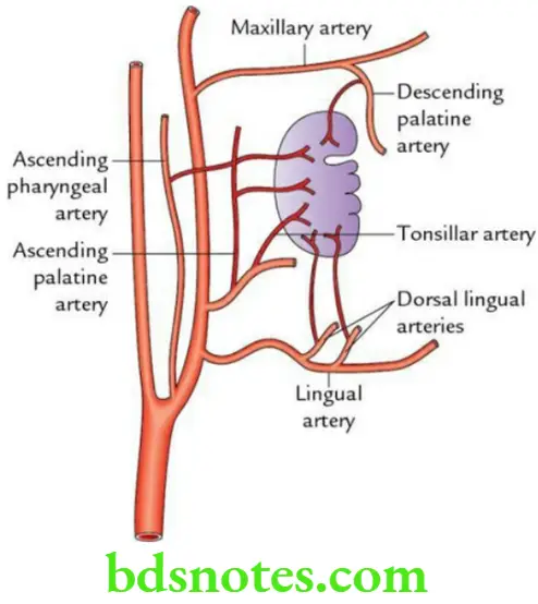
Arterial supply of the tonsil is supplied by the following five sets of arteries:
- Tonsillar branch of the facial artery (principal artery)
- Dorsal lingual branches of the lingual artery
- Ascending pharyngeal artery – a branch of the external carotid artery
- Ascending palatine artery – a branch of the facial artery
- Greater/descending palatine artery – a branch of the maxillary artery
Venous drainage: By peritonsillar vein into pharyngeal venous plexus, which in turn drains into the internal jugular vein.
Hard Soft Palate
Lymphatic drainage: Lymph vessels from the tonsil drain into jugulodigastric lymph nodes. These lymph nodes lie in the angle formed between posterior belly of digastric (inferior border) and internal jugular vein (anterior aspect) deep to the mandible.
“Comprehensive Overview Of Tonsils And Their Significance In Health”
Lymphatic drainage Applied anatomy
- Tonsillitis: It is an infection of the tonsil, which is usually of viral origin. This leads to the enlargement of jugulodigastric lymph nodes.
- Quinsy (peritonsillar abscess): It is the name given to collection of pus in the peritonsillar space.
- Referred pain: Pain of the tonsil is referred to middle ear because both are supplied by the glossopharyngeal nerve.
- Commonest source of bleeding after tonsillectomy. It is due to damage of the paratonsillar vein.
- After tonsillectomy, all blood clots in the tonsillar fossa are removed to prevent bleeding as removal of these clots allows the retraction of blood vessels due to muscle contraction. The only other organ in the body where such removal of blood clots is done is the uterus.
Anatomy And Physiology Of Tonsils
Question 1. What is gag reflex?
Answer.
It is a protective reflex characterized by elevation of the palate and contraction of pharyngeal muscles with associated retching and gagging in response to stimulation of mucosa of the oropharynx. The af erent limb of this reflex is formed by the glossopharyngeal nerve, while its ef erent limb is formed by the vagus nerve.
Question 2. Write a short note on the development of tonsil.
Answer.
The tonsil develops from the 2nd pharyngeal pouch in the 4th week of intrauterine life.
- The epithelial lining of the tonsil develops from the endoderm of the 2nd pharyngeal pouch.
- The stroma of tonsil develops from local mesenchyme.
- The lymphocytes of the tonsil are derived from either local mesenchyme or from circulating lymphocytes.
Classification Of Features On Dorsal Surface Of Tongue
“Best Practices For Diagnosing And Treating Tonsil-Related Disorders”
Question 1. Define tongue and list its functions.
Answer.
The tongue is a mobile, muscular organ situated on the floor of the mouth. It performs the following functions:
- Taste
- Speech
- Mastication
- Deglutition
“How To Live A Healthier Life By Supporting Tonsil Health”
Question 2. Enumerate the external features of the tongue.
Answer.
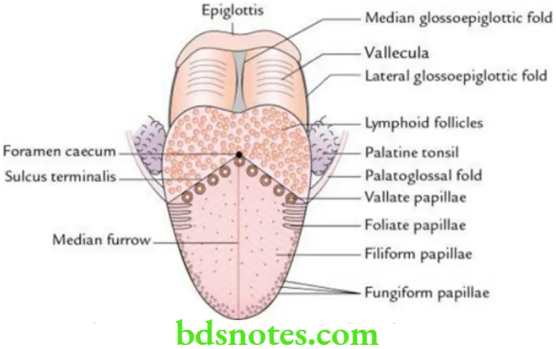
- The tongue has an apex, tip, and body.
- Body presents:
- Dorsal surface (also called dorsum)
- Ventral surface
- Right lateral margin
- Left lateral margin
Dorsum of Tongue
- Anatomically and developmentally, the dorsum of the tongue is divided into two parts: anterior two-thirds (oral part) and posterior one-third (pharyngeal part). The two parts are separated from each other by a V-shaped sulcus – the sulcus terminalis.
- A blind foramen at the apex of sulcus is called foramen caecum. The foramen caecum represents the site of development of the endodermal thyroglossal duct which grows down into the neck during embryonic development.
Features on the dorsal surface of Tongue
- Posterior one-third presents:
- A large number of lymphoid follicles, which together form lingual tonsils.
- A large number of openings of mucous and serous glands.
- Anterior two-thirds presents:
- A median furrow.
- A large number of papillae.
Features on the ventral surface of the tongue The ventral surface of the tongue presents:
- Frenulum linguae: A median fold of mucous membrane extending between the tongue and the floor of the mouth.
- Plica fimbriata: Two, fringed corrugated folds of mucous membrane, one on either side of frenulum linguae converging towards the tip of the tongue.
- Prominences of deep lingual veins: These are visible, one on either side, between frenulum linguae and plica fimbriata.
“The Role Of The Glossopharyngeal Nerve In Tonsil Innervation”
Anatomy And Histology Of The Types Of Tonsils
Discuss the histological features of a tonsil.
Answer.
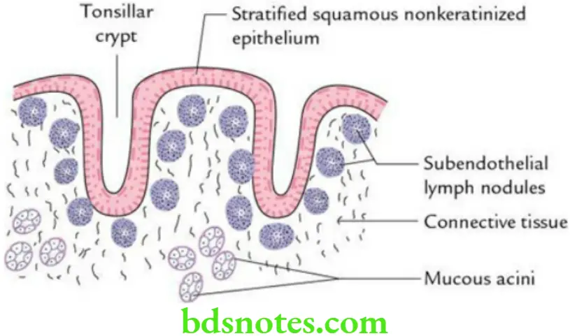
The histological features of the tonsil are as follows:
- The surface is lined by stratified, squamous, nonkeratinized epithelium.
- Surface epithelium dips at places into the substance of tonsil to form tonsillary crypts.
- Presence of subendothelial lymph nodules underneath the stratified squamous epithelium, along the crypts.
- Presence of mucous glands in the deeper part.
- Fibrous capsule on the outer side.
Leave a Reply