Submandibular Gland Excision
Describe the submandibular gland under the following headings:
- Submandibular Gland Location and parts,
- Submandibular Gland External features,
- Submandibular Gland Relations,
- Submandibular Gland Nerve supply and
- Submandibular Gland Applied anatomy.
Answer.
“Importance Of Early Treatment For Submandibular Gland Issues”
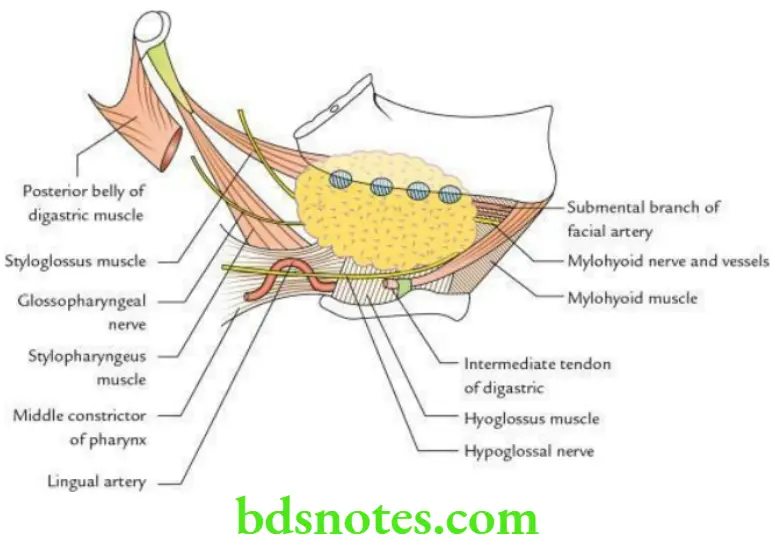
Submandibular Gland Location and Parts The submandibular gland lies in the digastric triangle. It is divided into two parts:
- Superficial
- Deep
The large, superficial part is located below the mylohyoid muscle and almost fills the digastric triangle.
The small, deep part is located above the mylohyoid muscle.
The two parts are continuous with each other around the free posterior margin of the mylohyoid muscle.
“Risk Factors For Complications During Submandibular Gland Excision”

“Early Signs Of Problems After Submandibular Gland Excision”
The gland is enclosed between the two layers of the investing layer of deep cervical fascia. The superficial layer covers the superficial surface of the gland and is attached to the base of the mandible. The deep layer covers the medial surface of the gland and is attached to the mylohyoid line of the mandible.
Superficial part of submandibular gland External features: It presents:
- Two ends: Anterior and posterior
- Three surfaces: Inferior, lateral and medial
Superficial part of submandibular gland Relations

“Understanding The Role Of Imaging In Submandibular Gland Excision”
Superficial part of submandibular gland Inferior surface is related to:
- Skin
- Superficial fascia
- Platysma
- Deep fascia
- Common facial vein
- Cervical branch of the facial nerve
- Submandibular lymph nodes
Superficial part of submandibular gland Lateral surface is related to:
- Submandibular fossa of mandible
- Medial pterygoid muscle
- Facial artery
Superficial part of submandibular gland Medial surface: It is extensive and is divided into three parts: anterior, intermediate and posterior.
- Anterior part is related to:
- Mylohyoid muscle
- Mylohyoid nerve and vessels
- Submental artery (a branch of facial artery)
- The intermediate part is related to:
- Hyoglossus muscle
- Lingual nerve
- Submandibular ganglion
- Hypoglossal nerve
- Submandibular duct
- The posterior part is related to:
- Styloglossus muscle
- Stylopharyngeus muscle
- Middle constrictor of the pharynx
- Glossopharyngeal nerve
- Lingual artery
“Step-By-Step Guide To Submandibular Gland Excision Surgery”
Deep part of the submandibular gland
- It is small and lies on the hyoglossus muscle.
- Posteriorly, it is continuous with a superficial part around the posterior border of the mylohyoid muscle.
- The submandibular duct emerges from its anterior end.
Nerve supply
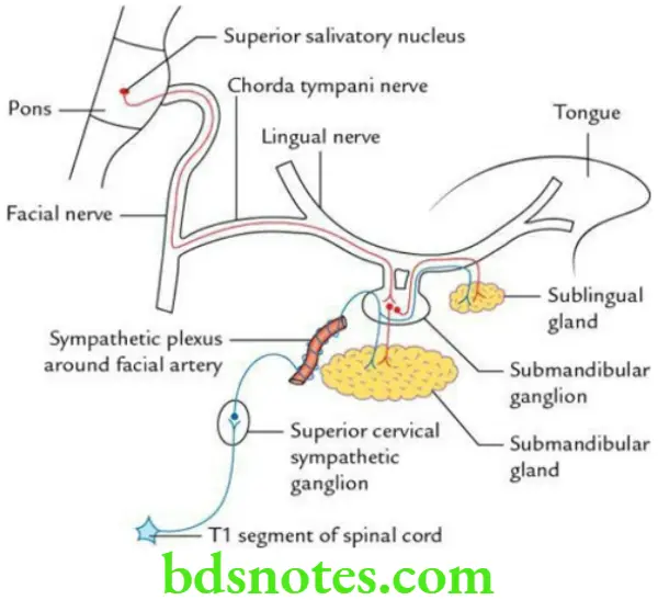
Secretomotor (parasympathetic):
- Preganglionic fibres arise from the superior salivatory nucleus and pass successively through the facial nerve, geniculate ganglion, chorda tympani and the lingual nerves to reach and relay in the submandibular ganglion.
- Postganglionic fibres arise from the submandibular ganglion and enter the submandibular gland to supply it.
Vasomotor (sympathetic):
- Preganglionic fibres arise from the T1 spinal segment and relay in the superior cervical sympathetic ganglion.
- Postganglionic fibres arise from the superior cervical sympathetic ganglion and run along the arteries to supply the gland. These fibres do not relay in the ganglion.
“The Role Of Minimally Invasive Techniques In Submandibular Gland Excision”
Sensory:
- Lingual nerve.
- Sensory fibres also do not relay in the ganglion.
Superficial part of submandibular gland Applied anatomy
- Formation of calculi is more common in the submandibular gland than in the parotid gland, because of (a) its secretion being more viscous and (b) the tortuous upward course of its duct (i.e. drainage occurs against gravity). This leads to stasis of secretion which leads to the formation of stone.
- To excise the submandibular gland, the skin incision is given at about 4 cm below the angle of the mandible to avoid injury to the marginal mandibular nerve.
- A stone in the submandibular duct can be palpated bimanually on the floor of the mouth and may even be seen if sufficiently large.
- Submandibular gland swelling can be palpated bimanually as it lies on both aspects of the oral diaphragm (i.e. mylohyoid muscle).
Leave a Reply