Spinal Nerves: Anatomy, Roots And Function
Briefly describe a typical spinal nerve. What is its distribution and applied anatomy?
Answer.
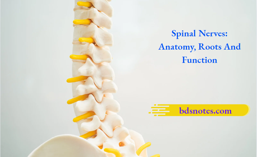
There are 31 pairs of spinal nerves. However, only T3 to T6 spinal nerves represent the typical spinal nerves.
The typical spinal nerve arises from the spinal cord by two roots: anterior and posterior. The anterior root is motor while the posterior root is sensory and possesses a ganglion called dorsal/posterior root ganglion.
“Importance Of Spinal Nerves In Transmitting Signals”

“Risk Factors For Damage To Spinal Nerve Roots”
The two roots unite to form the nerve trunk, which divides into two rami: dorsal and ventral. The small dorsal ramus passes backward to the muscles on the back of the vertebral column. Here, it divides into medial and lateral branches which supply the muscles. Thereafter, one of them divides into medial and lateral cutaneous branches to supply the overlying skin of the back of the body.
“Early Signs Of Issues With Spinal Nerve Function”
- Soon after its formation, the large ventral ramus is connected to the sympathetic ganglion by two rami communicantes – grey and white.
- Grey ramus communicans: It consists of nonmyelinated nerve fibers, which arise from the sympathetic ganglion. These fibers (sympathetic fibers) are distributed to the blood vessels, hair, and sweat glands through the branches of both the ventral and dorsal rami of the spinal nerve.
“Comprehensive Overview Of Spinal Nerves And Their Significance”
- White ramus communicans: It consists of myelinated nerve fibers, which arise from the lateral grey horn of the spinal cord and leave the ventral ramus to enter into the ganglion of the sympathetic trunk for its distribution.
- The large ventral ramus runs laterally into the intercostal space and supplies the intercostal muscles of this space. It also gives rise to lateral and anterior cutaneous branches.
- The cutaneous branches of the dorsal and ventral rami of the spinal nerve together supply a strip of skin from the posterior median line to the anterior median line called the dermatome.
“Understanding The Role Of Dorsal And Ventral Roots In Spinal Nerves”
Applied anatomy
- Spinal neuralgia: It is a sharp burning sensation in the area of skin supplied by the single spinal nerve (i.e. dermatome). The most important cause of this neuralgia is Herpes Zoster, a viral infection of the spinal ganglia.
- Root pain: Compression of nerve roots during their exit from intervertebral foramina in cases of spondylitis and disc prolapse causes pain along the distribution of that nerve.
Leave a Reply