Secondary Pulmonary Tuberculosis
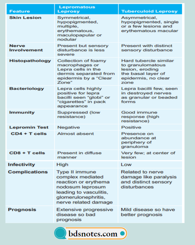
Question 1. Write the difference between tuberculoid and lepromatous leprosy.
Or
Write the differences between lepromatous and tuberculoid leprosy.
Answer:
Question 2. Write briefly about secondary pulmonary tuberculosis.
Answer:
The infection of an individual who has been previously infected or sensitized is called as secondary or post-primary or reinfection or chronic tuberculosis.
Secondary pulmonary tuberculosis
Secondary Pulmonary Tuberculosis
The lesions in secondary pulmonary tuberculosis usually begin as 1-2 cm apical area of consolidation of lung, which may in time develop a small area of central caseation necrosis and peripheral firosis.
It occurs by the hematogenous spread of infections from primary complex to the apex of affcted lung where the oxygen tension is high and favorable for growth of aerobic tubercle bacilli.
Fate of Secondary Pulmonary Tuberculosis
- The subapical lesion may heal with fibrous scaring and calcification.
- The lesions may coalesce together to form larger area of tuberculosis:
- Fibrocaseous tuberculosis
- Tuberculous caseous pneumonia
- Miliary tuberculosis.
- Tuberculous empyema.
Fibrocaseous Tuberculosis
The original area of tuberculosis pneumonia undergoes massive central caseation necrosis:
- The tubercular cavity is spherical with thick fibrous wall, lined by yellowish, caseous, necrotic material and the lumen is traversed by thrombosed blood vessels.
- Around the wall of cavity foci of consolidation are seen.
- The overlying pleura may also be thickened.
Microscopically
The wall of the cavity shows eosinophilic, granular, caseous material which may show foci of dystrophic calcification.
- Tubercular granulomas consist of epithelioid cells, Langhans giant cells and peripheral mantle of lymphocytes and central caseation necrosis.
- The outer wall of the cavity shows firosis.
Complications
- May produce hemoptysis
- Extending to pleura produces bronchopleural fitula
- Tubercular empyema
- Thickened pleura.
Tuberculosis upper lobe involvement
Tuberculous Caseous Pneumonia
In an individual with high degree of hypersensitivity, second- ary pulmonary tuberculosis may spread to rest of the lung, producing caseous pneumonia.
Miliary Tuberculosis
This is the lymphohematogenous spread of tuberculous infection.
- The spread may occur to systemic organ or isolated organs.
- The miliary lesions are millet seed sized (1 mm diameter), yellowish, fim areas.
- Lesions show the structure of tubercles with a minute area of caseation necrosis.
- The spread may be extrapulmonary into liver, spleen, kidney, brain and bone marrow.
Tuberculous Empyema
The caseating pulmonary lesions of tuberculosis may be associated with pleurisy as a reaction and is expressed as a series of fibrinous exudates.
Pleural effusion may heal by fibrosis and obliterate the pleural space. Occasionally the pleural cavity may contain caseous material and develop into tuberculous empyema.
Leave a Reply