Retinal Artery Occlusion
“Why is early detection of retinal artery occlusion critical for saving vision? Answered”
Retinal Artery Occlusion is the first and most important branch of the ophthalmic artery. It pierces the optic nerve 1.25 cm behind the eyeball reaches the optic disc through the central part of the disc and divides into four branches, one for each quadrant. These branches are superior nasal, inferior nasal, superior temporal and inferior temporal. It supplies the optic nerve and the inner six or seven layers of the retina.
Central Retinal Artery Applied Anatomy: Any blockage of a central retinal artery leads to loss of vision.
“Understanding the causes and symptoms of retinal artery occlusion through FAQs: Q&A explained”
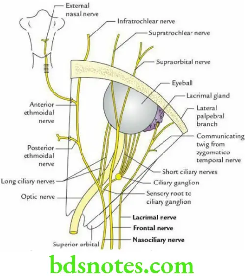
Origin, Course And Distribution Of Ophthalmic Nerve
- The ophthalmic nerve is the first and smallest division of the trigeminal nerve. It arises from the trigeminal ganglion and enters the lateral wall of the cavernous sinus. Here it divides into three branches, nasociliary, frontal and lacrimal, which enter the orbit through the superior orbital fissure.
- The nasociliary nerve runs forward and medially crosses the optic nerve obliquely from the lateral to the medial side, then runs along the medial wall of orbit to terminate by dividing into anterior ethmoidal and infratrochlear nerves.
It gives rise to the following branches:- Sensory root to the ciliary ganglion
- Two or three long ciliary nerves
- Posterior ethmoidal nerve
- Anterior ethmoidal nerve
- Infratrochlear nerve
“Importance of studying retinal artery occlusion for ophthalmologists and medical students: Questions explained”
- The frontal nerve (largest branch) runs forward and terminates by dividing into supraorbital and supratrochlear nerves.
- The lacrimal nerve (smallest branch) runs along the lateral wall of the orbit end and ends in the lacrimal gland.
It gives rise to the following branches:- Branches to the lacrimal gland
- The lateral palpebral branch to the skin of the lateral part of the upper eyelid
“Common challenges in diagnosing and treating retinal artery occlusion effectively: FAQs provided”
Leave a Reply