Production Of X-Ray
Question 1. describe in detail components of X-ray tube and functions of each component.
or
Draw labeled diagram of X-ray tube. (Main X-ray generating system).
or
Write short answer on components of an X-ray tube.
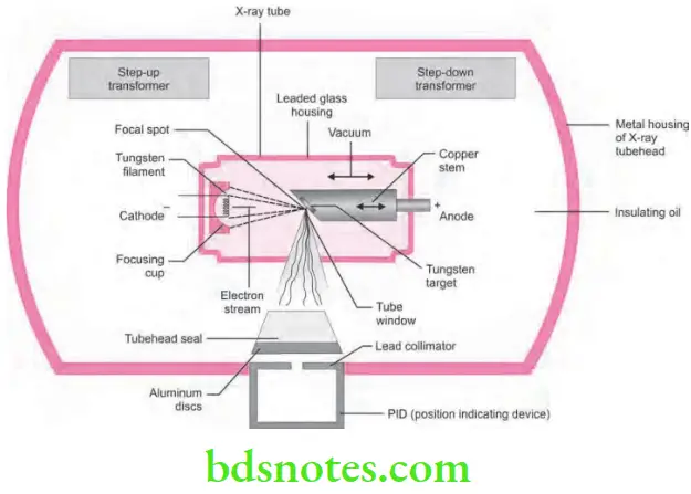
Answer. X ray tube is the heart of the X-ray generating system. This consists of a glass vacuum tube from which all of the air has been removed. The X-ray tube used in dentistry measures approximately several inches long by one inch in diameter.
Read And Learn More: Oral Radiology Question And Answers
“Understanding the role of X-ray production in medical imaging: Q&A explained”
The Parts of the X-ray tube
- A leaded glass housing
- A negative cathode
- A positive anode.
- A leaded glass housing: It is a leaded glass vacuum tube that prevents X-rays from escaping in all directions (radiation leakage). One central area of the leaded glass tube has a “window” that permits the X-ray beam to exit the tube and directs the X-ray beam towards the aluminum disk, lead collimator and position indicating device. This is also used for earthing.
- A negative cathode: It is principally composed of two parts:
- Filament.
- Focusing cup
“Importance of studying X-ray production for better imaging outcomes: Questions explained”
X-ray Tube Filament
- The filament is the source of electrons in the tube, it is made up of a coil of tungsten wire, approximately 0.2 cm in diameter, 1–2 mm wide, 0.1–0.2 mm thick and 7 to 15 mm in length. lt is mounted on two strong stiff wires, that support it and carry the electric current.
- These two mounted wires lead through the glass envelope to serve as a connection to the low and high voltage electrical source.
- The filament is heated to incandescence through a range of temperatures by varying voltage (10 V), across the filament from a step-down transformer in a low voltage circuit.
- The hot filament emits electrons that are separated from the outer orbits of tungsten atoms at a rate proportional to its temperature by a process called “Thermionic emission”.
“Common challenges in explaining X-ray production effectively: FAQs provided”
“Why is X-ray production critical for accurate diagnostics? Answered”
- The electrons lost by the filament form a cloud or space charge around the filament.
- Amilliampere control provides forfineadjustment of voltage across the filament and in turn the flow of heating current through it.
- The milliampere control, thereby controls the quantity of electrons the filament emits, which in turn controls tube current. Vaporization of the filament occurs over a period of time.
“Steps to explain the principles of X-ray production: Electron beam vs target interaction: Q&A guide”
- When the particles vaporize (turn into gaseous form) they solidify on the glass of the X-ray tube, which is called ’sunburning’ or ’sun-tanning of the tube. This reduces the output of the X-ray tube, destruction of the vacuum and integrity of the tube, resulting in ’arcing’ and ultimate tube failure. Thorium is added to the filament material to make the tube last longer.
X-ray Tube Focusing Cup
- The focusing cup is a negatively charged concave reflector cup of molybdenum or nickel and houses the filament.
- The focusing cup electrostatically focuses the electrons emitted by the incandescent filament into a narrow beam, directed at a small rectangular area in the anode—the focal spot.
- The electrons are caused to move in this direction because of a strong electric field interposed between the cathode and the anode, by a high negative charge placed on the cathode which repels the electrons in the electron cloud towards the anode which has a high positive charge. This is achieved by applying a high voltage circuit between the anode and the cathode.
“Role of the anode and cathode in X-ray production: Questions answered”
- To facilitate the movement of the electron cloud, the X—ray tube is evacuated as completely as possible to prelude collision of moving electrons with gas molecules, which could significantly reduce their speed. It also prevents oxidation or burn out of the filament.
- A positive anode or the positive electrode: lt consists of a wafer thin tungsten plate (target) embedded in a solid copper stem. The purpose of the target is to convert the kinetic energy of the electrons generated from the filament into X—ray photons. The position of the anode is indicated externally as a depression, usually a red dot on the tube head by the manufacturer.
“Early warning signs of issues addressed by proper X-ray production techniques: Common questions”
Question 2. Write short note on cathode in X-ray tube.
Answer. Cathode present in X-ray tube consists of negative electrode, so it is known as negative cathode.
Negative cathode Principally composed of two Parts
- Filament.
- Focusing cup
“Asymptomatic vs symptomatic effects of ignoring X-ray production protocols: Q&A”
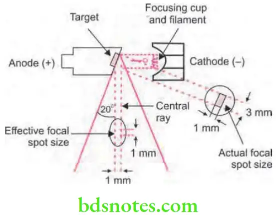
Question 3. Write short note on angle of truncation.
Answer.
“Role of counseling in clarifying X-ray production goals for patients: Questions answered”
In the practice, inclination of target is at 20° to central ray of electrons. This leads to effective focal spot to be 1 mm × 1 mm in contrast to 1 mm × 3 mm of actual focal spot size. This results in formation of small source of X-rays and sharp image with large actual focal spot for the effective heat dissipation. The 20° angle is known as angle of truncation.
Leave a Reply