The Primary Deciduous Teeth
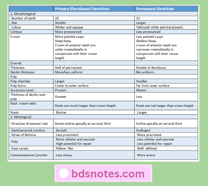
Question 1. Describe in detail the difference between deciduous (primary) dentition and permanent dentition.
Answer:
Read And Learn More: BDS Previous Examination Question And Answers
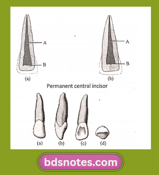
Question 2. Write about the Primary Maxillary Central Incisor.
Answer:
Primary Maxillary Central Incisor Aspects:
- Crown.
- Greater mesiodistal diameter than the cervicofacial length.
- Labial surface – smooth.
- Incisal edge- straight.
- Root length greater than crown length.
Primary Maxillary Central Incisor Lingual aspect:
- Marginal ridges – well developed.
- Cingulum
- Well developed.
- Extends up to the incisal ridge.
- Divides into mesial and distal fossa.
- Root narrows lingually.
- Cross-section- triangular in shape.
Primary Maxillary Central Incisor Mesial and Distal Aspects:
- Wider crown at cervical third.
- The curvature of the cervical third.
- The curvature of the cervical line is distinct and curves towards the incisal edge.
- Cervical curvature distally is less than the curvature medially.
- Roots appear blunter.
- The mesial surface of the root has concavity and the distal surface is convex.
Primary Maxillary Central Incisal Aspects:
- Straight incisal edge.
- The labial surface of it is broader and smoother.
- Proximal surfaces are broad.
Question 3. Describe the Primary Maxillary Canine.
Answer:
- Labial aspect.
- Constricted crown at the cervix.
- Mesial and distal surfaces are more convex.
- The incisal edge has a long, well-developed, sharp cusp.
- Mesial slope of the cusp is longer than the distal slope of the cusp.
- A line drawn through the contact areas of the deciduous canine would bisect a line drawn from the cervix to the tip of the cusp.
Primary Maxillary Canine Lingual aspect:
- It shows pronounced enamel ridges that merge with each other.
- It shows cingulum, mesial and distal marginal ridges, incisal cusp ridges, lingual ridge, and tubercle at the cusp tip.
- The lingual ridge divides the lingual surface into shallow mesiolingual and distolingual fossae the root of this tooth tapers lingually.
Primary Maxillary Canine Mesial aspect:
- The labiolingual measurement at the cervical third is much greater.
- It permits resistance against forces the tooth must withstand during function.
- The function of this tooth is to punch tear and apprehend food material.
Primary Maxillary Canine Distal aspect:
- The curvature of the cervical line toward the cusp ridge is less than the mesial surface.
Primary Maxillary Canine Incisal aspect:
- The Crown is diamond-shaped.
- Has more pronounced and less rounded lines and point angles.
- The tip of the cusp is distal to the center of the crown.
- The mesial cusp slope is longer than the distal cusp slope.
- This allows for intercuspation with the mandibular canine which has its longest slope distally.
Question 4. Describe about Primary Mandibular Central Incisor.
Answer:
Primary Mandibular Central Incisor Labial aspect:
- Has a flat face with no development grooves.
- Mesial and distal sides are tapered evenly.
- Has long and tapered roots.
- The root is almost twice the length of the crown.
Primary Mandibular Central Incisor Lingual aspect:
- The marginal ridges and cingulum are located.
- The middle third and incisal third have a flat surface or slightly concave forming. Lingual fossa.
- Crown and root converge lingually.
Primary Mandibular Central Incisor Mesial aspect:
- The incisal ridge is centered over the center of the root.
- The convexity of the cervical contours labially and lingually at the cervical third.
- The mesial surface of the root is nearly flat and evenly tapered.
Primary Mandibular Central Incisor Distal aspect:
- It is the reverse of the mesial aspect.
- The cervical line of the crown is less curved towards the incisal ridge.
- Developmental depression present over the distal side of the root.
Primary Mandibular Central Incisor Incisal aspect:
- The incisal ridge is straight.
- It bisects the crown labiolingually. Taper is seen towards the cingulum
- The labial surface presents a flat surface that is slightly convex.
- The lingual surface presents a flat surface that is slightly concave.
Question 5. Describe in detail about Primary Maxillary First Molar.
Answer:
Primary Maxillary First Molar Buccal Aspects:
- The maxillary first molar is the widest at the mesial and distal contact area.
- The crown converges at the cervix.
- The occlusal line is slightly scalloped. The buccal surface is smooth.
- Slight developmental grooves are noted.
- The roots are slender, long, and widely spread.
- The distal root is shorter than mesial
- The bifurcation of the roots begins almost immediately at the site of the cervical line.
Primary Maxillary First Molar Lingual aspects:
- The crown converges in a lingual direction.
- The mesiolingual cusp is the most prominent cusp, the longest and sharpest cusp.
- The distolingual cusp is poorly defined, small, and rounded.
- Distobuccal cusp is seen as longer and better developed.
- All three roots are seen with the lingual root being larger.
Primary Maxillary First Molar Mesial aspect:
- The dimension at the cervical third is greater than that at the occlusal third.
- Pronounced convexity is seen at the cervical third.
- The cervical line mesially shows some curvature in the occlusal direction.
- The mesiobuccal root is hidden behind the mesiobuccal root.
Primary Maxillary First Molar Occlusal aspect:
- The calibration of the distance between the mesiobuccal line angle and the distobuccal line angle is greater than the calibration between the mesiolingual line angle and the distolingual line angle.
- The crown converges lingually, distally.
- It is more nearly rectangular in shape with the shortest sides being the marginal ridges.
- A mesial triangular fossa is inside the mesial marginal ridge, with a mesial pit in this fossa and a sulcus with its central groove connecting the two fossae.
- Buccal development groove divides the mesiobuccal cusp and distobuccal cusp occlusally.
- Supplemental grooves radiate from the pit in the mesial triangular fossa one buccally, one lingually and one towards the marginal ridge.
- A well-developed triangular ridge called oblique ridge connects mesiolingual cusp with the distobuccal cusp.
- The central development groove extends from mesial pit to distal developmental groove.
- The distal marginal ridges is thin and poorly developed.
Question 6. Describe in distal about Primary Maxillary Second Molar.
Answer:
Primary Maxillary Second Molar Buccal aspect:
- It shows two well-defined buccal cusps with a buccal developmental groove between them.
- The crown is narrow at the cervix.
- The roots appear slender and longer.
- The point of bifurcation between the buccal roots is close to the cervical line.
- The two buccal cusps are nearly equal in size.
Primary Maxillary Second Molar Lingual aspect:
- It shows three cusps.
- Mesiolingual cusp – large.
- Distolingual – well-developed.
- Supplemental cusp=apical to mesiolingual cusp called tubercle of carabelli.
- It is poorly developed.
- It acts as a buttress to the bulk of the mesiolingual cusp.
- If it is missing, some traces of developmental lines or “dimples” remain.
- A well-defined development groove separates the mesiolingual cusp from the distolingual cusp.
- It connects with the developmental groove.
- All three roots are visible.
- The lingual root is large and thick.
Primary Maxillary Second Molar Mesial aspect:
- The mesiolingual cusp appears large.
- The mesiobuccal cusp is short and sharp.
- Little curvature to the cervical line is seen.
- The mesiobuccal root is broad and flat.
- The lingual root appears long and slender.
- The point of bifurcation between the mesiobuccal root and the lingual root is 2-3 mm apical to the cervical line of the crown.
- The mesiolingual cusp is below the bifurcation.
- Cervical curvature is lingual.
Primary Maxillary Second Molar Distal aspect:
- A lingual outline creates a smooth, rounded line.
- Distobuccal and distolingual cusp are of the same length.
- The cervical line is straight.
- All three roots are seen.
- The distobuccal root is shorter and narrower.
- The point of bifurcation between the distobuccal root and lingual root is more apical.
Primary Maxillary Second Molar Occlusal aspect:
- It is somewhat rhomboidal.
- It shows 4 cusps.
- Mesiobuccal.
- Distobuccal
- Mesiolingual
- Distolingual
- Supplemental cusps.
- The buccal surface is flat with a developmental groove.
- Occlusal surface has.
- A central fossa with a central pit.
- A well-defined mesial triangular fossa is distal to the mesial marginal ridge.
- The oblique ridge connects the mesiolingual and distobuccal cusp.
- Central groove connecting mesial triangular fossa with the central fossa.
- Buccal developmental groove.
- Extends buccally from the central pit.
- Separates triangular ridges.
- Lingual developmental groove.
- Separates mesiolingual and distolingual cusps.
- Supplemental grooves.
-
- Radiates from developmental grooves.
- Distal fossa- distal to oblique ridge.
- Distal triangular fossa.
- Mesial to the distal marginal ridge.
Question 7. Describe in detail about Primary Mandibular First Molar.
Answer:
Primary Mandibular First Molar Buccal aspect:
- It has a straight outline.
- The Crown constricts at the cervix.
- The distal portion of the crown is shorter than the mesial portion.
- Cervical line dip apically.
- The mesial cusp is larger than the distal cusp.
- Development depression divides them. The roots are long and slender
Primary Mandibular First Molar Lingual aspect:
- Crown and root converge lingually.
- The distolingual cusp is rounded.
- The mesiolingual cusp is long and sharp.
- Developmental groove occurs between the distolingual and mesiolingual cusp.
- The cervical line is straight.
Primary Mandibular First Molar Mesial aspect:
- Mesiobuccal and mesiolingual cusps are seen.
- The mesial marginal ridge is well-developed.
- The cervical line slants upward.
- The root end is flat and almost square.
Primary Mandibular First Molar Distal aspect:
- The cervical line is almost straight.
- The distal marginal ridge is not well defined.
- The distal root is rounder, shorter, and tapers apically.
Primary Mandibular First Molar Occlusal aspect:
- The mesiolingual cusp is the largest and most well-developed.
- The buccal developmental groove divides the two buccal cusps.
- The central developmental groove separates the mesiobuccal and mesiolingual cusps.
- Two supplemental grooves are seen.
- The mesiobuccal cusp exhibits a well-defined triangular ridge.
- Lingual developmental grooves separate media lingual and distolingual cusp.
Question 8. Describe in detail about Primary Mandibular Second Molar.
Answer:
Primary Mandibular Second Molar Buccal aspect:
- It is wider at the cervical portion.
- Mesiobuccal and distobuccal developmental grooves divide the buccal surface into mesiobuccal, buccal, and distobuccal cusp.
- Roots appear slender and long.
- Roots flare mesiodistally at the middle and apical third.
- The point of bifurcation of the roots is just below the CEJ.
Primary Mandibular Second Molar Lingual aspect:
- Two equal cusps with a lingual groove between them are seen.
- The cervical line is relatively straight.
- A portion of each of the three buccal cusps may be seen. Roots appear slender and long.
Primary Mandibular Second Molar Mesial aspects:
- The tooth seems constricted occlusal. The lingual cusp is longer.
- The cervical line is regular.
- The mesial root is broad and flat.
Primary Mandibular Second Molar Distal aspect:
- Mesiobuccal and distobuccal cusps are seen distolingual cusp appears well developed.
- The triangular ridge extends from the distolingual cusp tip to the distal marginal ridge.
- The cervical line of the crown is regular.
- The distal root is broad and flat and tapers more at the apical end.
Primary Mandibular Second Molar Occlusal aspect:
- It is somewhat rectangular.
- Buccal cusps are the same in size, while both lingual cusps are equal.
- Well-defined triangular ridges are seen.
- The occlusal surface shows.
- Mesial triangular fossa.
- Inside the mesial marginal ridge.
- Distal triangular fossa.
- Mesial to the distal marginal ridge.
- Central developmental groove.
- Extends from mesial triangular fossa to distal triangular fossa.
- Two buccal grooves-mesial and distal.
- Lingual developmental groove.
- Supplemental grooves.
- Seen over slopes of triangular ridges and in mesial and distal triangular fossae.
- The mesial marginal ridge is well developed.
- Crown converge distally.
Leave a Reply