Periodontal Ligament, Its Structure And Function
“What is the periodontal ligament and why is it important?”
The periodontal ligament is composed of a complex vascular and highly cellular connective tissue that surrounds the tooth root and connects it to the inner wall of the alveolar bone.
The periodontal ligament is the soft richly vascular and cellular connective tissue which surrounds the root of the teeth and joins the root cementum with the lamina dura or the alveolar bone proper or the socket wall.
Read And Learn More: Periodontics Question And Answers
“Understanding the role of the periodontal ligament in tooth stability”
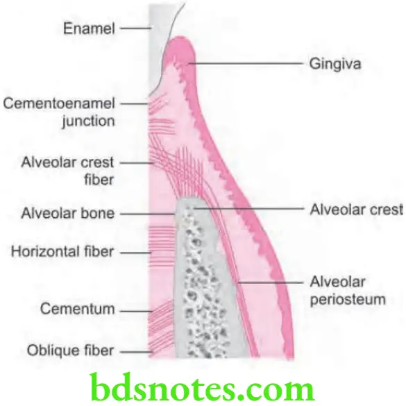
Structure of Periodontal Ligament
- Periodontal ligament is continuous with the connective tissue of the gingiva, and it communicates with the marrow spaces via vascular channels present inside the bone.
- Average width of periodontal ligament space is 0.2 mm, considerable variation exists.
- Periodontal space is diminished around teeth which are not in function and in unerupted teeth, but it get increased in teeth which have been subjected to hyperfunction.
- Periodontal ligament consists of the following:
- Periodontal fibers
- Principal fibers
- Secondary fibers
- Cellular elements
- Ground substance
- Periodontal fibers
“Importance of studying the periodontal ligament for dental professionals”
Periodontal Fibers
Principal Fibers of Periodontal Ligament
- Most important elements of the periodontal ligament are principal fibers, which are collagenous and arranged in bundles and follow a wavy course when viewed in longitudinal section.
- Terminal portions of the principal fibers which are inserted into cementum and bone are known as Sharpey fibers.
- Principal fiber bundles consist of individual fibers which form a continuous anastomosing network between tooth and bone.
- The principal fibers of the periodontal ligament are arranged in six groups that develop sequentially in the developing root i.e.
- Transseptal group
- Alveolar crest group
- Horizontal group
- Oblique group
- Apical group
- Interradicular group
“Common challenges in analyzing the periodontal ligament”
Principal Fibers Of The Periodontal Ligament – Transseptal group
- Transseptal fibers extend interproximally over the alveolar bone crest and get embedded in the cementum of adjacent teeth.
- These fibers are reconstructed even after destruction of the alveolar bone which results from periodontal disease.
- Transseptal fibers may be considered as belonging to the gingiva, because they do not have osseous attachment.
- These fibers lead to the maintenance of the teeth in the arch.
“Steps to identify layers of the periodontal ligament under a microscope”
Principal Fibers Of The Periodontal Ligament – Alveolar crest group
- They extend obliquely from cementum just beneath the junctional epithelium to the alveolar crest.
- These fibers also run from the cementum over the alveolar crest and to firous layer of the periosteum which covers the alveolar bone.
- Alveolar crest fibers prevent extrusion of tooth and resist lateral tooth movements.
- Incision of these fibers at the time of periodontal surgery does not increase tooth mobility unless significant attachment loss has occurred.
- These fibers secure teeth inside the socket by resisting the lateral forces which are applied to the tooth.
“Role of collagen fibers in the structure of the periodontal ligament”
Principal Fibers Of The Periodontal Ligament – Horizontal group
- These fibers extend at right angles to the long axis of tooth from cementum to the alveolar bone.
- These fibers mainly prevent the lateral tooth movement.
Principal Fibers Of The Periodontal Ligament – Oblique group
- Oblique fibers constitute the largest group in periodontal ligament and they extend from the cementum in a coronal direction obliquely to the bone.
- They absorb the chewing forces over the tooth and are the main support of the tooth.
- They resist the apically directed masticatory forces.
Principal Fibers Of The Periodontal Ligament – Apical group
- Apical fibers radiate in irregular manner from the cementum to the bone at the apical region of the socket.
- They do not occur on incompletely formed roots.
- They prevent the tooth tipping and resist the forces of luxation.
Principal Fibers Of The Periodontal Ligament- Interradicular group
- These fibers fan out from the cementum to the tooth in the furcation areas of multirooted teeth.
- They prevent tipping of tooth, forces of luxation and rotation.
“How does the periodontal ligament connect teeth to bone?”
Secondary Fibers of Periodontal Ligament
- Periodontal ligament has two immature forms are found: oxytalan and elaunin.
- The so-called oxytalan fibers run parallel to the root surface in a vertical direction and bend to attach cementum in cervical third of root. They are thought to regulate vascular low.
- Oxytalan fibers have been shown to develop de novo in the regenerated periodontal ligament.
- In addition to these fiber types, small collagen fibers associated with the larger principal collagen fibers have been described. Such fibers run in all directions and form a plexus called the indifferent fiber plexus.
Cellular Elements of the Periodontal Ligament
Four types of cells have been identified in the periodontal ligament:
- Connective tissue cells
- Epithelial rest cells
- Immune system cells
- Cells associated with neurovascular elements.
Connective Tissue Cells
- Connective tissue cells consist of firoblasts, cementoblasts and osteoblasts.
- Fibroblasts are most common cells in periodontal ligament and they appear as ovoid or elongated cells which are oriented along the principal fibers and exhibit pseudopodia-like processes.
- Fibroblasts synthesize collagen and have the capacity to phagocytose “old” collagen fibers and degrade them be the enzyme hydrolysis. So collagen turnover appears to be regulated by firoblasts.
- Osteoblasts, cementoblasts, osteoclasts, and odontoclasts are also seen in the cemental and osseous surfaces of the periodontal ligament.
“Early warning signs of abnormal periodontal ligament changes”
Epithelial Rests of Malassez
- These cells form a latticework in periodontal ligament and appear as either isolated clusters of cells or interlacing strands which depends on the plane in which the microscopic section is cut.
- Epithelial rests are considered to be the remnants of the Hertwig root sheath, which disintegrates during root development.
- Epithelial rests are distributed close to the cementum throughout the periodontal ligament of most teeth; they are most numerous in the apical area and cervical area.
- These cells diminish in number with age by degenerating and disappearing or by undergoing calcifiation to become cementicles.
- The cells are surrounded by a distinct basal lamina, they are interconnected by hemidesmosomes and they contain tonofiaments.
- Although the functional properties of these cells are still considered to be unclear but they are reported to contain keratinocyte growth factors, and they have been shown to be positive for tyrosine kinase A neurotrophin receptor. In addition, epithelial rests proliferate when stimulated and they participate in the formation of periapical cysts and lateral root cysts.
Defense Cells
- They are neutrophils, lymphocytes, macrophages, mast cells, and eosinophils.
- These cells and the cells which are associated with neurovascular elements, are similar to the cells which are found in other connective tissues.
Ground Substance
“Asymptomatic vs symptomatic stages of ligament damage”
- Periodontal ligament consists of a large proportion of ground substance that fills the spaces between fibers and cells.
- Ground substance consists of two main components i.e.
- Glycosaminoglycans, which are hyaluronic acid and proteoglycans
- Glycoproteins which are fironectin and laminin.
- Ground substance consists of high water content i.e.70%.
- Cell surface proteoglycans participate in several biologic functions, including cell adhesion, cell–cell and cell–matrix interactions, binding to various growth factors as coreceptors, and cell repair.
“Can targeted microscopy improve diagnostic accuracy?”
“Steps to teach students about the periodontal ligament”
Functions of Periodontal Ligament
Physical functions Periodontal Ligament
- Provision of soft tissue casing: For protecting the vessels as well as nerves from injury by the mechanical forces, periodontal ligament provides the soft tissue casing around them.
- Transmission of occlusal forces to bone:
- Axial force when applied to tooth, it causes stretching of oblique fibers of periodontal ligament. Transmission of this tensional force to the alveolar bone encourages bone formation rather than bone resorption.
- But when horizontal or tipping force is applied the tooth rotates around the axis, at fist the tooth movement is within the confies of the periodontal ligament. When a greater force is applied, displacement of facial and lingual plates may occur.
- Apical portion of root moves in the in direction opposite to coronal portion. In areas of tension, principle fibers of bundle are taut rather than wavy. In areas of pressure, fibers get compressed, tooth gets displaced and corresponding distortion of bone occur in direction of root movement.
- The axis of rotation, in single-rooted teeth is located in the area between the apical and middle third of the root. Root apex and coronal half of the clinical root are suggested as other locations of axis of rotation.
- In multirooted teeth, the axis of rotation is located at the furcation area.
- Resistant to impact of occlusal forces: Two Theories have been explained for the mechanism of tooth support:
- Tensional theory: According to it, the principal fibers of periodontal ligament plays a major role in supporting the tooth and transmittng forces to the bone. When forces are applied to the tooth, principal fibers unfold and straighten and then transmit the forces to the alveolar bone, causing elastic deformation of the socket. Finally when the alveolar bone has reached its limit, the load is transmittd to basal bone.
- Viscoelastic theory is based on the fact that, the fluid movement largely controls the displacement of the tooth, with fibers playing a secondary role. When the forces are transmittd to the tooth, the extracellular fluid is pushed from the periodontal ligament into the marrow spaces through the cribriform plate. after the depletion of tissue fluids, the bundle fibers absorb the shock and tighten. This leads to blood vessel stenosis followed by arterial back pressure followed by ballooning of the vessels and replenishing with tissue fluids.
- Maintains the gingival tissues in their proper relationship to the teeth.
- Shock absorption resists the impact of occlusal forces.
“Can interactive tools improve learning outcomes?”
Formative and Remodeling Function
- Periodontal ligament and alveolar bone cells are exposed to physical forces in response to mastication, parafunction, speech, and the orthodontic tooth movement.
- Cells of periodontal ligament participate in formation and resorption of cementum and the bone, which occur at the time of physiologic tooth movement, during the accommodation of periodontium to occlusal forces, and during the repair of injuries.
- Periodontal ligament shows variations in cellular enzyme activity which are correlated with the remodeling process. Although applied loads may induce vascular and inflammatory reactive changes in periodontal ligament cells.
“Role of diagrams and models in explaining ligament layers”
- The periodontal ligament is constantly undergoing remodeling. Old cells and fibers are broken down and replaced by new ones, and mitotic activity may be observed in the firoblasts and endothelial cells.
- Fibroblasts form the collagen fibers, and the residual Mesenchymal cells develop into osteoblasts and cementoblasts. Therefore the rate of formation and the differentiation of osteoblasts, cementoblasts, and fibroblasts affect the rate of formation of collagen, cementum, and bone.
Nutritional and Sensory Functions
- Periodontal ligament supplies nutrients to the cementum, bone and gingiva via blood vessels, and it also provides lymphatic drainage. As compared to other ligaments and tendons, the periodontal ligament is highly vascularized tissue. High blood vessel content may provide hydrodynamic damping to applied forces as well as high perfusion rates to the periodontal ligament.
- Periodontal ligament is richly supplied with sensory nerve fibers which are capable of transmittng tactile, pressure, and pain sensations through trigeminal pathways. Nerve bundles pass in the periodontal ligament from the periapical area and through channels from the alveolar bone that follows the course of the blood vessels.
- The bundles divide into single myelinated fibers, which loose their myelin sheaths and end in one of four types of neural termination i.e.
“Early warning signs of knowledge gaps in ligament education”
- Free endings, which have a treelike confiuration and carry pain sensation
- Ruffini-like mechanoreceptors, which are located primarily in the apical area
- Coiled Meissner’s corpuscles and mechanoreceptors, which are found in mid – root region
- Spindle-like pressure and vibration endings, which are surrounded by a fibrous capsule and located mainly in apex.
Leave a Reply