Periodontal Flap Surgery
Following are the flap techniques:
- Modified Widman flap.
- Undisplaced flap.
- Palatal flap.
- Apically displaced flap.
- Distomolar surgery.
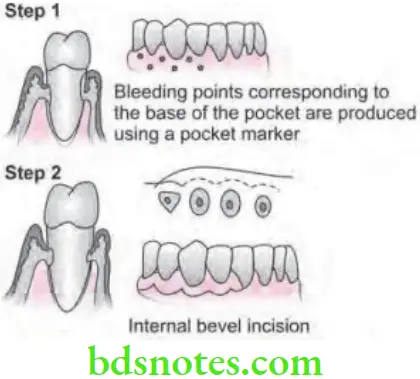
Undisplaced Flap
In this procedure, the entire soft tissue pocket wall is removed with initial incision.
“Techniques Used In Periodontal Flap Surgery”


“Procedures Involved In Periodontal Flap Surgery”
- Step 1: Pockets are measured with periodontal probe and bleeding point is produced on the outer surface of gingiva to mark the base of the pocket. In this procedure, final placement of flap is determined by first incision.
- Step 2: Initially, internal bevel incision is made following the scalloping bleeding points made on the gingiva, incision is usually carried to a point apical to alveolar crest depending on thickness of tissue. The thicker the tissue, more apical will be the end point of the incision.
- Step 3: The second or crevicular incision is made from the bottom of pocket to bone to detach connective tissue from bone.
- Step 4: Flap is then reflected with periosteal elevator.
- Step 5: Interdental incision is made with a interdental knife.
- Step 6: Triangular wedge of tissue is created by three incisions removed with a curette.
- Step 7: Area is debrided, removing tissue tags and granulation tissue with sharp curette. The roots are scaled.
- Step 8: Flap is then placed back to end at root bone junction.
- Step 9: Flaps are sutured together with continuous sling suture or interrupted sutures.
“Best Ways To Understand Periodontal Flap Surgery”
Flap Surgery: Types, Procedure & Recovery
Indications for Periodontal Flap Surgery
- In gaining the access for root debridement.
- For reducing or eliminating the pocket depth, so that patient can maintain root surfaces free of plaque.
- For reshaping soft and hard tissues to attain harmonious topography.
- For regenerating the alveolar bone, periodontal ligament and cementum.
“Indications For Periodontal Flap Surgery In Dentistry”
Contraindications for Periodontal Flap Surgery
- In patients having poor plaque control.
- In patients with high caries rate.
- In unrealistic patient expectation and desire.
- In uncontrolled medical conditions such as:
“Risk Factors For Complications In Periodontal Flap Surgery”
- Unstable angina
- Uncontrolled diabetes
- Uncontrolled hypertension
- Myocardial infarction/Stroke within the six months.
Leave a Reply