Norma Occipitalis
Question 1. External occipital protuberance
Answer:
External occipital protuberance Site:
- External occipital protuberance is an median prominence in the lower part of norma occipitalis
External occipital protuberance Significance:
- External occipital protuberance marks the junction of the head and neck
- The most prominent part of it is the inion
- External occipital protuberance is easily felt at the point where the back of the neck becomes continuous with the scalp
External occipital protuberance Attachments:
- Upper part
- Gives origin to the trapezius
External occipital protuberance Lower part:
- Gives attachment to the upper end of the ligamentum nuchae.
“What is norma occipitalis? A detailed question and answers guide”
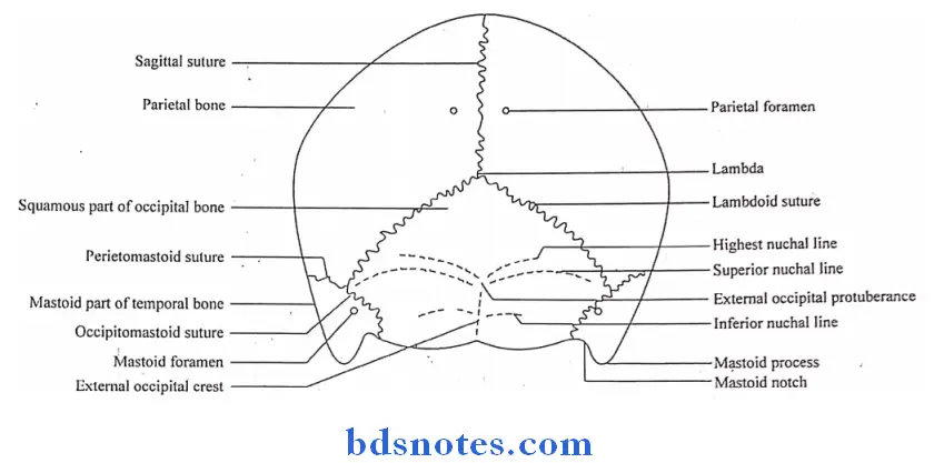
Question 2. Superior nuchal lines
Answer:
- Superior nuchal lines are curved bony ridges passing laterally from the external occipital protuberance
Superior nuchal lines Attachments:
- Medial one-third: gives origin to trapezius
- Lateral part: provides insertion to sternocleidomastoid above and splenius capitis below
“Understanding norma occipitalis and norma lateralis through FAQs: Anatomy, structures, and uses explained”
Question 3. Superciliary arch
Answer:
Superciliary arch Location & Appearance:
- It is a rounded, curved elevation situated just above the medial part of each orbit
- Prominent in males
Superciliary arch Attachments:
- Medial part: gives origin to the corrugators supercilli
Normal Lateralis
Question 1. Mastoid process.
Answer:
- The mastoid process is a nipple-like large projection from the lower part of the mastoid temporal bone
- The mastoid process forms the lateral wall of the mastoid notch
Mastoid process Location:
- Posteroinferior to the external acoustic meatus
Mastoid process Occurrence:
- During the 2nd year of life
Mastoid process Structures Present:
- Tympanomastoid fissure: on the anterior aspect of the base of mastoid process
- Mastoid foramen: at near occipitomastoid suture
Mastoid process Structures Attached:
- Sternocleidomastoid, splenius capitus & longissimus capitus inserted from before backwards on the posterior part of the lateral surface of the mastoid process
- Posterior belly of digastrics – from mastoid notch
Mastoid process Relations:
1. Tympanomastoid fissure
- Transmits auricular branch of vagus nerve
2. Mastoid foramen – transmits
- Emissary vein connecting the sigmoid sinus with the posterior auricular vein
- Meningeal branch of occipital artery
“Importance of studying norma occipitalis and norma lateralis for medical students: Questions explained”
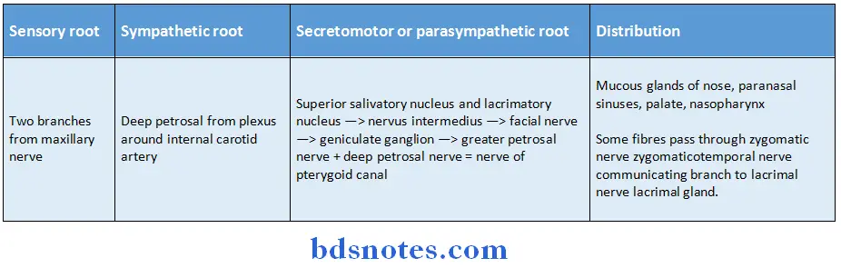
Position, Connection And Branches Of Pterygopalatine Ganglion
Pterygopalatine Ganglion Situation:
Pterygopalatine Ganglion is the largest parasympathetic ganglion
Pterygopalatine Ganglion Situation Branches:
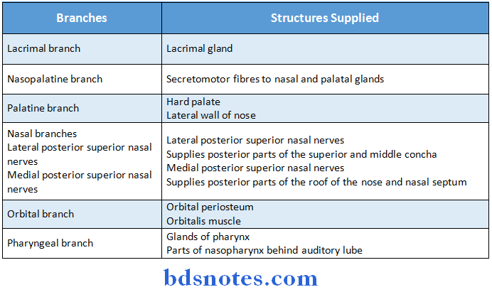
Pterygopalatine Ganglion Situation Connections:
“Common challenges in mastering norma occipitalis and norma lateralis notes effectively: FAQs provided”
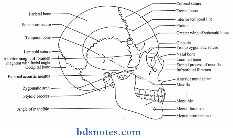
Zygomatic arch
- The zygomatic arch is the horizontal bar
Zygomatic arch Location:
- Sympathetic root
Zygomatic arch Formation:
- Anterior 1/3rd: by the temporal process of the zygomatic bone
- Posterior 2/3rd: by the zygomatic process of the temporal bone
Zygomatic arch Parts:
- Jugal point: At the anterior end of the upper border
- Anterior & posterior root: At the posterior end
- Articular tubercle: At the junction of anterior & posterior root
- Postglenoid tubercle: Behind the articular fossa
Zygomatic arch Attachments:
Temporal fascia: Outer & inner parts of superior border of arch
Masseter: To the medial & lower border of arch
Lateral ligament of TMJ: To the tubercle of root of the arch
“Why is identifying norma occipitalis critical for occipital surgeries? Answered”
Pterion
- Pterion is an H-shaped suture present in the anterior part of the floor of the temporal fossa
- The pterion is a meeting point of four bones
- Frontal
- Parietal
- Greater wing of sphenoid
- Temporal
Pterion Location:
- 4 cm above the midpoint of zygoma
- 2.5 cm behind the frontozygomatic suture
Pterion Relations:
- Structures related to it are:
- Middle meningeal vein
- Anterior division of the middle meningeal artery
- Stem of the lateral sulcus of the brain
Bones Meeting At Pterion
- Frontal
- Parietal
- Greater wing of sphenoid
- Temporal
Surgical importance of pterion
- Pterion is the thin part of skull
- In roadside accidents, the anterior division of the middle meningeal artery may be ruptured, leading to clot formation between the skull bone & the dura mater or extradural haemorrhage
Result:
- Clot compresses the motor area of the brain, leading to paralysis of the opposite side
Prevention: Use of a helmet
“Factors influencing success with skull base studies: Q&A”
Mastoid process
- Mastoid Process is a nipple-like large projection from the lower part of the mastoid temporal bone
- Mastoid Process forms the lateral wall of mastoid notch
Mastoid Process Location:
- Posteroinferior to the external acoustic meatus
Mastoid Process Occurrence:
- During the 2nd year of life
Structures Present:
Tympanomastoid fissure: On the anterior aspect of the base of mastoid process
Mastoid foramen: At/ near occipitomastoid suture
Mastoid Process Structures Attached:
- Sternocleidomastoid, splenius capitus and longissimus capitus- inserted from before backwards on the posterior part of the lateral surface of the mastoid process
- Posterior belly of digastrics – from mastoid notch
“Steps to explain structures of norma occipitalis: Occipital bone vs foramen magnum vs lambdoid suture: Q&A guide”
Muscles Attached To Mastoid Process
Sternocleidomastoid, splenius capitus and longissimus capitus- inserted from before backwards on the posterior part of the lateral surface of the mastoid process
- Posterior belly of digastrics – from mastoid notch
Mastoid Groove
- The mastoid part of the temporal bone lies just behind the external acoustic meatus
- It is continuous with the squamous temporal bone
- A partially obliterated squamomastoid suture may be visible just in front of & parallel to the roughened area of muscular insertions known as the mastoid groove.
Stylomastoid Foramen Location:
- Stylomastoid Foramen is located posterior to the styloid process
Stylomastoid Foramen Structures passing through it:
- Facial nerve
- Stylomastoid branch of posterior auricular foramen
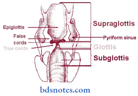
Piriform Fossa/ pṣiriform recess
- Piriform Fossa is a small cavity or pocket between the lateral walls of the pharynx on each side & upper part of the larynx
- Piriform Fossa is also called Pyriform fossa or Pyriform sinus
“Role of the occipital bone in protecting the brainstem: Questions answered”
Suprameatal triangle of McEwan’s
- Suprameatal triangle is a small depression present posteroinferior to the external auditory meatus.
- McEwans forms the lateral wall of the tympanic or mastoid antrum.
McEwans Boundaries:
- Superior – supramastoid crest
- Anterior – posterior superior margin of the external auditory meatus
- Posterior – vertical tangent to the posterior margin of the meatus.
Leave a Reply