Norma Basalis
“What is norma basalis? A detailed question and answers guide”
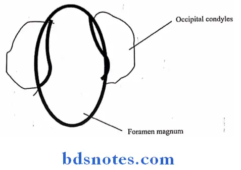
Foramen magnum
- Foramen magnum is largest foramen of the skull
- Opens into posterior cranial fossa-upwards and into vertebral canal- downward
- Foramen magnum is oval in shape
- Foramen magnum is overlapped on each side by the occipital condyles
Foramen magnum Structures Passing Through It:
- Anterior part-3 structures
- Apical ligament of dens
- Vertical band of cruciate ligament
- Membrana tectoria
- Posterior part-2 structures
- Lower part of medulla oblongata
- 3 meninges
- Through subarachnoid space- 9 Structures
- Spinal accessory nerves -2
- Vertebral arteries – 2
- Sympathetic plexus around the vertebral arteries -2
- Posterior spinal arteries – 2
- Anterior spinal artery – 1
“Understanding norma basalis through FAQs: Anatomy, structures, and uses explained”
Structures passing through foramen magnum
- Anterior part-3 structures
- Apical ligament of dens
- Vertical band of cruciate ligament
- Membrana tectoria
- Posterior part-2 structures
- Lower part of medulla oblongata
- 3 meninges
- Through subarachnoid space-9 Structures
- Spinal accessory nerves -2
- Vertebral arteries – 2
- Sympathetic plexus around the vertebral arteries -2
- Posterior spinal arteries – 2
- Anterior spinal artery – 1
“Importance of studying norma basalis for medical students: Questions explained”
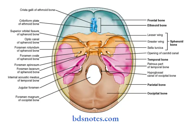
Foramen of middle cranial fossa
“Common challenges in mastering norma basalis notes effectively: FAQs provided”
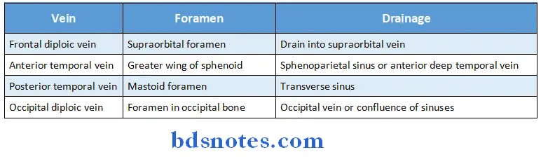
Diploic veins
- The diploic veins are large, thin-walled valveless veins that channel in the diploë between the inner and outer layers of the cortical bone in the skull.
- They develop fully by the age of two years.
- The diploic veins drain this area into the dural venous sinuses.
- The four major types of diploic veins found on each side of the head are frontal, anterior temporal, posterior temporal, and occipital diploic veins.
Types:
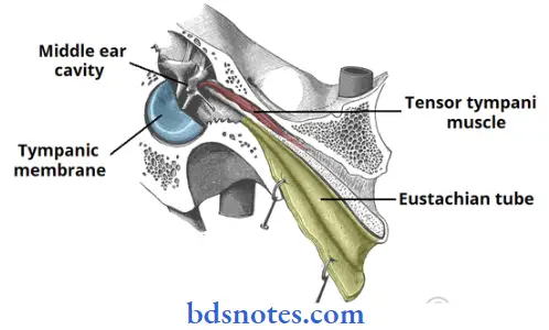
Auditory tube
- The auditory tube connects the middle ear with the nasopharynx anteriorly
- The sulcus tube which is a groove between the posteromedial margin of the greater wing of the sphenoid & the petrous temporal bone lodges the cartilaginous part of the auditory tube
Pterygomaxillary fissure
- Pterygomaxillary fissure is a triangular interval formed by the divergence of the maxilla from the pterygoid process of the sphenoid
- Pterygomaxillary fissure connects infratemporal with the Pterygopalatine fossa
- Pterygomaxillary fissure transmits terminal part of internal maxillary artery
“Why is identifying norma basalis critical for skull base surgery? Answered”
Pterygopalatine Fossa
Pterygopalatine Fossa Location:
- Lies deep to the pterygomaxillary fissure
Pterygopalatine Fossa Boundaries:
- Anterior
- Posterior surface of maxilla
- Posterior
- Pterygoid process
- Greater wing of sphenoid
- Medial
- Perpendicular place of palatine bone
- Floor
- Union of anterior & posterior walls
Pterygopalatine Fossa Contents:
- Maxillary nerve & its branches
- 3rd part of maxillary artery & its branches
- Pterygopalatine ganglion with its branches
“Factors influencing success with norma basalis studies: Q&A”
Contents Of Pterygopalatine Fossa
3 contents:
- Maxillary nerve & its branches
- 3rd part of maxillary artery & its branches
- Pterygopalatine ganglion with its branches
3 ganglion:
- Sphenopalatine
- Pterygopalatine
- Ganglion of hay fever
3 structures transversing in posterior wall:
- Maxillary nerve
- Nerve of pterygoid canal
- Pharyngeal branch through pterygoid canal
3 structures passing through inferior orbital fissure
- Infraorbital nerve
- Zygomatic nerve
- Orbital branches of the ganglion
3 structures through inferior opening:
- Anterior palatine nerve & greater palatine vessels
- 2 Posterior palatine nerve & lesser palatine vessels
3 structures through medial opening:
- Nasopalatine nerve & sphenopalatine vessels
- Medial posterosuperior nasal branches
- Lateral posterosuperior nasal branches
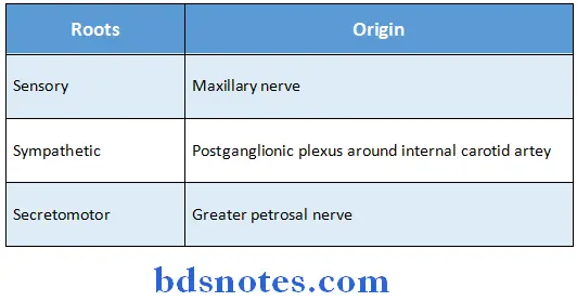
3 roots of the ganglion:
- Sensory
- Sympathetic
- Secretomotor
“Steps to explain clinical relevance of norma basalis: Surgical planning vs trauma management vs imaging techniques: Q&A guide”
Pterygopalatine ganglion
- Pterygopalatine ganglion is largest parasympathetic ganglion
Pterygopalatine ganglion Roots
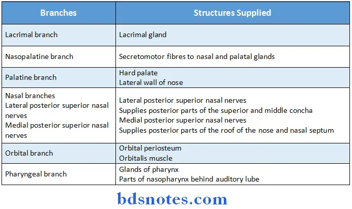
Pterygopalatine ganglion Branches
“Role of norma basalis in guiding surgical approaches: Questions answered”
- Posteriorly, the groove leads to the bony part of the auditory tube, which lies within the petrous temporal bone
Pterygopalatine ganglion Parts
- Osseous part-36 mm long
- Fibro-cartilaginous part-24 mm long
Pterygopalatine ganglion Arterial Supply:
- Middle meningeal artery
- Artery of the pterygoid canal
- Ascending pharyngeal branch of external carotid artery
Pterygopalatine ganglion Venous Drainage:
- Pterygoid & pharyngeal Venous plexus
Pterygopalatine ganglion Function:
- Maintains equilibrium of air pressure
Foramen magnum
- Foramen magnum is largest foramen of the skull
- Opens into posterior cranial fossa-upwards and into vertebral canal- downward
- Foramen magnum is oval in shape
- Foramen magnum is overlapped on each side by the occipital condyles
“How does trauma to norma basalis affect cranial nerves? FAQ explained”
Jugular foramen
- Jugular foramen is large & elongated.
- Jugular foramen is placed at the posterior end of the petro-occipital suture
Jugular foramen Structures passing through it:
- Anterior part:
- Inferior petrosal sinus
- Meningeal branch of ascending pharyngeal artery
- Middle part:
- Glossopharyngeal nerve
- Vagus nerve
- Accessory nerve
- Posterior part:
- Internal jugular vein
- Meningeal branch of occipital artery
“Early warning signs of poor adherence to skull base principles: Common questions”
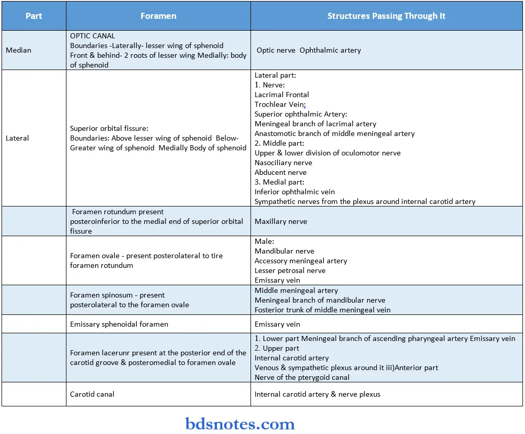
Foramen lacerum
- Foramen lacerum is short, wide canal, 1 cm long
- Bounded by
- Posterolaterally: by apex of the petrous temporal bone
- Medially:
- Basiocciput
- Body of the sphenoid
- Anteriorly
- Root of the pterygoid process
- Greater wing of the sphenoid bone
Foramen lacerum Structures passing through it:
- Lower part
- Meningeal branch of ascending pharyngeal artery
- Emissary vein
- Upper part
- Internal carotid artery
- Venous & sympathetic plexuses around it
- Anterior part
- Nerve of the pterygoid canal
Superior orbital fissure
- Superior orbital fissure is situated at the posterior part of the junction between the roof & lateral wall of the orbit
- Superior orbital fissure is an oblique, roughly triangular space bounded
- Above by the lesser wing
- Below by the greater wing
- Medially by the body of the sphenoid
Structures passing through it:
- Lateral part:
- Nerves: lacrimal, frontal, trochlear
- Vein: superior ophthalmic
- Arteries: meningeal branch of lacrimal artery, anastomotic branch of middle meningeal artery
- Middle part:
- Upper & lower division of oculumotor nerve
- Nasociliary nerve
- Abducent nerve
- Medial part:
- Inferior ophthalmic vein
- Sympathetic nerves from the plexus around internal carotid artery
“Asymptomatic vs symptomatic effects of delayed interventions: Answered”
Structures passing through foramen ovale
Male:
- Mandibular nerve
- Accessory meningeal artery
- Lesser petrosal nerve
- Emissary vein
Structures passing through foramen spinosum
Structures passing through foramen spinosum are:
- Middle meningeal artery
- Meningeal branch of mandibular nerve
- Posterior trunk of middle meningeal vein
“Can preventive measures reduce risks of skull base fractures? FAQs provided”
Structures passing through superior orbital fissure
- Lateral part
- Nerves: lacrimal, frontal, trochlear
- Veins: superior ophthalmic
- Arteries: meningeal branch of lacrimal artery, anastomotic branch of middle meningeal artery
- Middle part
- Upper and lower division of oculomotor nerve
- Nasociliary nerve
- Abducent nerve
- Medial part
- Inferior ophthalmic vein
- Sympathetic nerves from the plexus around internal carotid artery
“Differential applications of static vs dynamic assessment tools: Questions answered”
Structures passing through anterior condylar canal
- Hypoglossal nerve
- Meningeal branch of the hypoglossal nerve
- Meningeal branch of ascending pharyngeal artery
- Emissary vein
Foramen ovale
- Foramen ovale is large and oval in shape
Foramen ovale Location:
Posterolateral to the upper end of the posterior border of lateral pterygoid plate
Foramen ovale Structures passing through it:
Male:
- Mandibular nerve
- Accessory meningeal artery
- Lesser petrosal nerve
- Emissary vein
“Role of better surgical outcomes in improving patient satisfaction: Questions answered”
Leave a Reply