Nasal Cavity: Anatomy, Structure, Parts, Blood Supply
Describe the Nasal Septum under the following headings:
- Nasal Septum Formation,
- Nasal Septum Arterial supply,
- Nasal Septum Nerve supply and
- Nasal Septum Applied anatomy.
Answer.
“Early Signs Of Issues With The Nasal Cavity”
Nasal Septum Formation
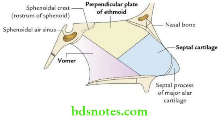
The nasal septum is a median osseocartilaginous partition between two nasal cavities covered on each side by the mucous membrane.
- The bony part is formed by:
- Vomer, below and behind
- The perpendicular plate of the ethmoid, above
- The cartilaginous part is formed by:
- Septal cartilage
- Septal processes of major alar cartilages
- The particular part is formed by:
- Fibrofatty tissue
“How To Care For Patients With Nasal Cavity Issues”
“Understanding The Role Of The Nasal Cavity In Respiratory Function”
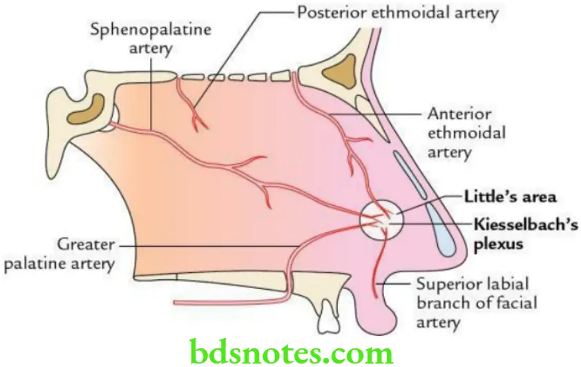
Nasal Septum Arterial supply
- Anterosuperior part, by anterior ethmoidal artery
- Posteroinferior part, by sphenopalatine artery
- Anteroinferior part, by superior labial and greater palatine arteries
- Posterosuperior part, by sphenopalatine artery
Nasal Septum Nerve supply
General sensory:
- Anterosuperior part by internal nasal branches of the anterior ethmoidal nerve
- Anteroinferior part by anterior superior alveolar nerve
- Posterosuperior part by medial, posterior, and superior nasal branches of the pterygopalatine ganglion
- Posteroinferior part by nasopalatine nerve – a branch of the pterygopalatine ganglion
“Comprehensive Overview Of The Nasal Cavity And Its Significance”
Special Sensory By The Olfactory Nerve.
Nasal Septum Applied anatomy
Deviated nasal septum (DNS): It may occur as a sequel to postnasal trauma (the most common cause) or due to congenital malformation. Excessive deviation of the nasal septum may cause nasal obstruction. It is treated by submucous resection (SMR) of the septum.
Epistaxis: It is nose bleeding that commonly occurs due to trauma of Kiesselbach’s plexus in the Little’s area.
“The Role Of Imaging In Diagnosing Nasal Cavity Disorders”
Question 1. What is the nose? List its functions.
Answer.
The nose is a pyramidal-shaped projection in the midface. It presents the tip (apex), alae, dorsum, root, and nostrils or nares. Its cavity is divided into two halves by a median nasal septum. Each cavity (also called the nasal cavity) communicates anteriorly to the exterior through the nostril (anterior nare) and posteriorly with the nasopharynx through the choana (posterior nare).
Nose Functions
- Is the organ of smell.
- Plays a significant role in respiration.
- Provides protection to the lower respiratory tract.
- Performs air conditioning of inspired air.
- Provides vocal resonance to voice.
Question 2. Enumerate the bones and cartilages forming the skeleton of external nose.
Answer.
Bones (four in number)
- Two nasal bones
- Frontal processes of maxillae
Cartilages (five in number)
- Two lateral nasal cartilages/superior nasal cartilages
- A single, median septal cartilage
- Two major alar cartilages/inferior nasal cartilages
Facial Vein: Anatomy, Tributaries, Drainage
Write a short note on the dangerous area of the face.
Answer.
Dangerous area of the face: This includes:
- Upper lip
- The lower part of the nose including the nasal septum
- Adjoining parts of the cheek
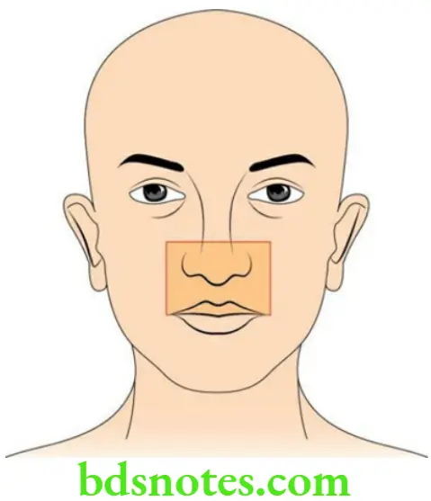
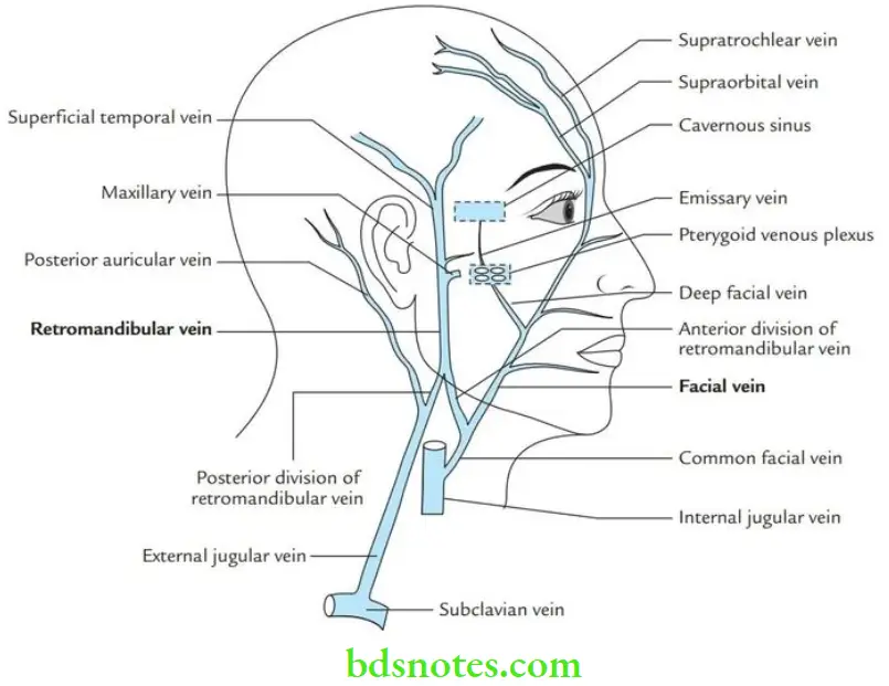
This area is dangerous because infective emboli from this area can reach the cavernous sinus and cause cavernous sinus thrombosis. As a result, cranial nerves present within the cavernous sinus are compressed leading to paralysis of the muscles of the eyeball.
Route of spread: The venous blood from the dangerous area of the face is drained into the cavernous sinus as follows:
Deep facial vein → pterygoid venous plexus → emissary vein → cavernous sinus
“Best Practices For Diagnosing And Treating Nasal Cavity Issues”
Paranasal Sinuses: Anatomy, Function And Types
Write briefly about frontal air sinuses.
Answer.
- These are located in the frontal bone between its outer and inner tables behind superciliary arches. The right and left frontal air sinuses are rarely of equal size, and the septum separating them is rarely situated in the median plane.
- The frontal sinus drains through the frontonasal duct inferiorly into a funnel-shaped infundibulum at the anterior end of the hiatus semilunaris of the middle meatus.
- The infection of the frontal sinus (frontal sinusitis) usually causes severe and localized pain in the forehead (frontal headache). The frontal headache shows characteristic periodicity, i.e. it increases as the sun rises and decreases as the sun sets.
- The pain of the frontal air sinus may extend up to the vertex through the supraorbital nerves that supply it. The frontal sinusitis can lead to brain abscess in the frontal lobe.
Epistaxis: Practice Essentials, Anatomy, Pathophysiology
What is Little’s area? Describe its clinical importance.
Answer.
It is an area in the anteroinferior part of the nasal septum where four arteries anastomose form an arterial plexus called Kiesselbach’s plexus.
The arteries forming this plexus are as follows:
- Septal branch of sphenopalatine
- Septal branch of the greater palatine
- Septal branch of anterior ethmoidal artery
- Septal branch of the superior labial artery – a branch of the facial artery
Kiesselbach’s Plexus Clinical importance The Little’s area is the most common site of nose bleeding (i.e. epistaxis) in young adults, usually due to fingernail trauma (nose picking)/small ulcer. The septal branch of the sphenopalatine artery is the largest, longest, and most tortuous. It is the main source of bleeding. Hence, it is also termed the artery of nose bleeding/rhinologist’s artery.
Leave a Reply