Middle Ear – Explanation, Structure, Anatomy, And Functions
Describe the middle ear under the following headings:
- Middle Ear Location, shape, and dimension,
- Middle Ear Contents,
- Middle Ear Boundaries,
- Middle Ear Nerve supply and
- Middle Ear Applied anatomy.
Answer.
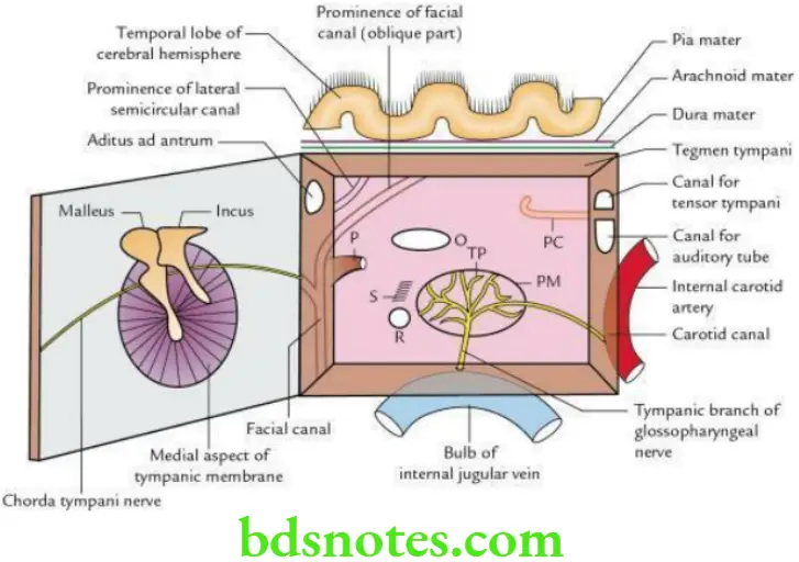
Middle Ear Location
- The middle ear is a narrow, slit-like, air-filled cavity in the petrous part of the temporal bone.
- Middle Ear communicates anteriorly with the nasopharynx through the auditory tube and posteriorly with the mastoid antrum through the aditus ad antrum.
“Early Signs Of Issues With The Middle Ear”
Middle Ear Shape Biconcave hollow disc, resembling the red blood cell (RBC).
Middle Ear Dimension
Vertical: 15 mm
Middle Ear Transverse:
- 6 mm: At roof
- 2 mm: In the center
- 4 mm: At floor
“Role Of The Ossicles In Sound Transmission”
Middle Ear Contents
- Air
- Two muscles: Tensor tympani and stapedius
- Three ear ossicles: Malleus, incus and stapes
- Two nerves: Chorda tympani nerve and tympanic plexus.
Middle Ear Boundaries
Middle Ear Roof or tegmental wall:
Formed by tegmen tympani, a thin plate of bone that separates the middle ear from the middle cranial fossa.
“Understanding The Role Of The Middle Ear In Sound Transmission”
Middle Ear Floor or jugular wall
- Formed by the jugular fossa of the temporal bone.
- Separates the middle ear from the superior bulb of the internal jugular vein.
Middle Ear Anterior Wall Or Carotid Wall
- The upper part presents Canals for tensor tympani and auditory tube.
- Lower part forms: Posterior wall of carotid canal.
“Best Practices For Diagnosing And Treating Middle Ear Infections”
Middle Ear Medial Or Labyrinthine Wall
Middle Ear separates the middle ear from the internal ear and presents the following features:
- Promontory: A rounded elevation produced by the first turn of the cochlea.
- Oval window (fenestra vestibuli): An oval opening posterosuperior to the promontory that leads to the vestibule of the internal ear. It is occupied by the base of stapes.
- Round window (fenestra cochleae): A round opening posteroinferior to the promontory that leads to the scala tympani of the cochlea and is closed by a secondary tympanic membrane.
- Sinus tympani: A depression behind promontory.
- The prominence of the facial canal is just above the oval window.
- The prominence of the lateral semicircular canal is above the prominence of the facial canal.
Middle Ear Lateral Or Membranous Wall
- Separates the middle ear from the external ear.
- It is formed:
- Mainly by the tympanic membrane.
- Partly by the squamous part of the temporal bone in the region of the epitympanic recess.
“Step-By-Step Guide To Explaining The Anatomy Of The Middle Ear”
Middle Ear Posterior Wall Or Mastoid Wall
It presents the following features. From above downwards, these are:
- Aditus to mastoid antrum (aditus ad antrum)
- Fossa incudis: Depression for incus.
- Pyramid: A conical bony projection. The apex of the pyramid presents an opening for the tendon of the stapedius.
- Posterior canaliculus for chorda tympani, lateral to pyramid.
Middle Ear Nerve Supply
- Tympanic branch of glossopharyngeal nerve.
- Superior and inferior caroticotympanic nerves from sympathetic plexus around the internal carotid artery.
“Tips To Prevent Damage To The Middle Ear Effectively”
Middle Ear Applied Anatomy
Otitis media:
The throat infections commonly spread to the middle ear through the auditory tube. It is more common in children because in children the tube is shorter, wider, and horizontal.
The longstanding otitis media often leads to a collection of pus in the middle ear – a condition called chronic suppurative otitis media (CSOM). The pus from the middle ear:
“The Role Of Imaging In Diagnosing Middle Ear Disorders”
- May be discharged in the external ear following the rupture of the tympanic membrane.
- May erode the roof leading to meningitis and temporal lobe abscess.
- May erode the floor causing thrombosis of the internal jugular vein.
- May spread backward into the mastoid antrum leading to a mastoid abscess.
Bleeding from ear: Fracture of the middle cranial fossa can cause bleeding through the ear.
Leave a Reply