• Skip to main content
  • Skip to secondary menu
  • Skip to primary sidebar
  • About Us
  • Terms of Use
  • Privacy Policy
  • Disclaimer
  • Contact Us
  • Sitemap

BDS Notes

BDS notes, Question and Answers

  • Public Health Dentistry
  • Periodontics
  • Pharmacology
  • Pathology
  • Orthodontics
    • Anchorage In Orthodontics
    • Mandibular Growth, Functional Matrix
    • Retention and Relapse
  • General Surgery
    • Cysts: Types, Causes, Symptoms
    • Maxillofacial Fractures, Disorders, and Treatments
    • Lymphatic Disorders
    • Neurological and Facial Disorders
  • Temporal And Infratemporal Regions
    • Spinal and Neuroanatomy
  • Dental Materials
    • Dental Amalgam
Home » Medial Medullary Syndrome and Pons Anatomy: Key Structures and Clinical Correlates

Medial Medullary Syndrome and Pons Anatomy: Key Structures and Clinical Correlates

November 16, 2025 by Joankessler parkland Leave a Comment

Medial Medullary Syndrome and Pons Anatomy: Key Structures and Clinical Correlates

Question 1. Write a short note on medial medullary syndrome.

Answer.  Causes Of Medial Medullary Syndrome 

Ischaemia of the medial region of the medulla due to thrombosis of the anterior spinal artery.

medial medullary syndrome

Clinical Features Of Medial Medullary Syndrome 

  • Contralateral hemiplegia (UMN type of paralysis) due to involvement of (pyramidal tract)
  • Ipsilateral paralysis of the tongue due to the involvement of the hypoglossal nerve (LMN type of paralysis)
  • Contralateral loss of proprioceptive sensations due to involvement of the medial meniscus

Pons

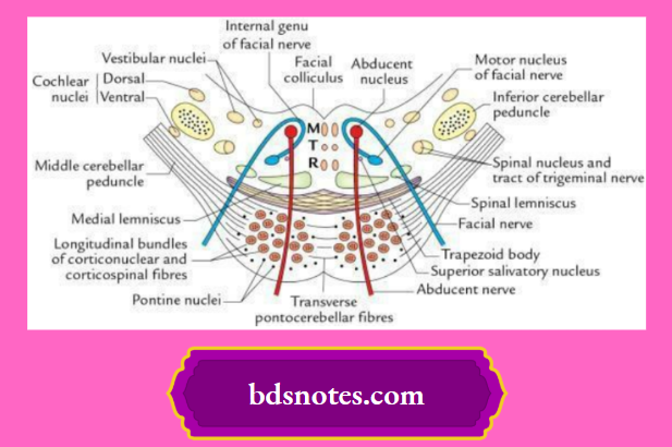
Question 2. Enumerate the main structures seen in the transverse section of the pons at the level of the facial colliculi.

The main structures seen in the transverse section of the pons at the level of the facial colliculi are as follows:

pons anatomy

  • Abducent nucleus
  • Motor nucleus of the facial nerve
  • Internal genu of the facial nerve

cranial nerve nuclei in pons

  • Dorsal and ventral cochlear nuclei
  • Trapezoid body

Brainstem Cranial Nerve Nuclei

Filed Under: Brain

Reader Interactions

Leave a Reply Cancel reply

Your email address will not be published. Required fields are marked *

Primary Sidebar

Recent Posts

  • Brainstem Anatomy Explained: Parts, Functions, and Clinical Significance
  • Medulla Oblongata Transverse Sections: Key Structures at Decussation Levels
  • Transverse Section of Medulla Oblongata: Upper Olive Level Structures Explained
  • Medial Medullary Syndrome and Pons Anatomy: Key Structures and Clinical Correlates
  • Upper Pons Cross‑Section: Trigeminal Nuclei and Lemnisci Unpacked
  • Midbrain Cross-Section at the Inferior Colliculus: Key Structures Explained
  • Medial Longitudinal Fasciculus (MLF): Anatomy, Pathways, and Functions
  • Cerebellum Simplified: Parts and Anatomical Lobes Explained
  • Cerebellar Nuclei Explained: Dentate, Interposed, and Fastigial Connections
  • Cerebellar Nuclei And Tracts – Structure, Connections And Functions
  • The Fourth Ventricle
  • Rhomboid Fossa
  • Central sulcus
  • Lobes Of The Brain
  • The Anatomy Of The Cerebral Cortex
  • Brodmann Area 4
  • Wernicke’s Area
  • Visual Cortex
  • Cerebral Dominance In Spatial Hearing
  • Paracentral Lobule

Copyright © 2025 · Magazine Pro on Genesis Framework · WordPress · Log in