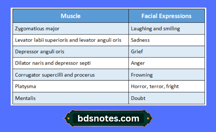
Platysma
“What are the layers of the scalp? A detailed question and answers guide”
- Platysma is one of the muscle of neck
Platysma Origin:
- Upper part of pectoral & deltoid fascia
- Fibres run upwards and medially
Platysma Insertion:
- Anterior fibres, to the base of the mandible
- Posterior fibres to the skin of the lower face and lip & may be continuous with the risorius
Platysma Action:
- Releases pressure of skin on the subjacent veins
- Depresses mandible
- Pulls the angle of the mouth downwards as in horror or surprise
“Understanding the layers of the scalp through FAQs: Composition, functions, and uses explained”
Platysma Nerve Supply:
- Cervical branch of facial nerve
Platysma Examination:
- Forcible pulling of the angles of the mouth downwards & backward forming prominent vertical folds of skin on the side of neck
Facial expression associated with It:
- Horror, terror, fright
“How do the layers of the scalp contribute to protection and function? FAQ answered”
Zygomatic Major
Zygomatic major Origin:
- From the surface of the zygomatic bone lateral to zygomatic minor muscle
Facial expression associated with it: Smiling & laughing
Nerve Supply: Buccal branch of facial nerve
SCALP mnemonic question and answer
“Importance of studying the layers of the scalp for medical students: Questions explained”
Orbicularis Oris
Orbicularis oris Parts:
1. Intrinsic part
- It is very thin sheet
Origin: - Superior incisivus from maxilla
- Inferior incisivus from mandible
Insertion: - Angle of mouth
2. Extrinsic part.
- It has two strata formed by converging muscles
Origin: - Thickest middle stratum from buccinators
- Thick superficial stratum from elevator & depressor of lip & their angles
Insertion: - Lips & the angle of the mouth
Action: - Closes & purses the mouth
Nerve Supply: - Temporal branch of facial nerve
Examination: - Whistling & pursing the mouth
“Common challenges in mastering layers of the scalp notes effectively: FAQs provided”
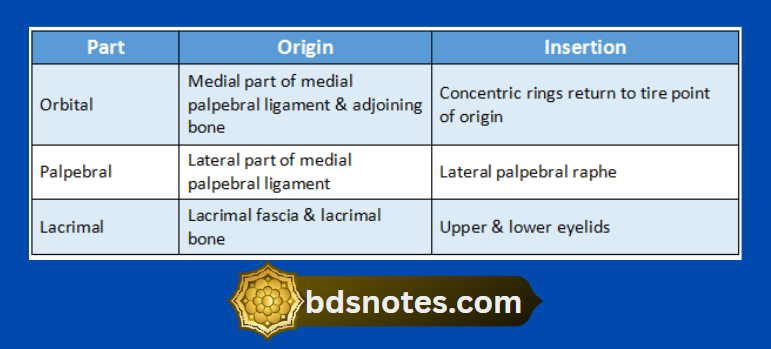
Orbicularis Oculi
Orbicularis oculi Parts:
“Why is proper understanding of the layers of the scalp critical for diagnosing scalp disorders? Answered”
Orbicularis oculi Actions:
1. Orbital part
- It closes the eye tightly, wrinkling
- Protects eye from bright light
2. Palpebral part
- Closes the eye gently in sleep or in blinking
3. Lacrimal part
- Draws the lacrimal papillae medially
- Dilates lacrimal sac
- Supports lower lid
Orbicularis oculi Examination:
- Tight closure of the eyes
Orbicularis oculi Applied Anatomy:
- Paralysis of this muscle leads to
- Drooping of the lower eyelid
- Spilling of tears
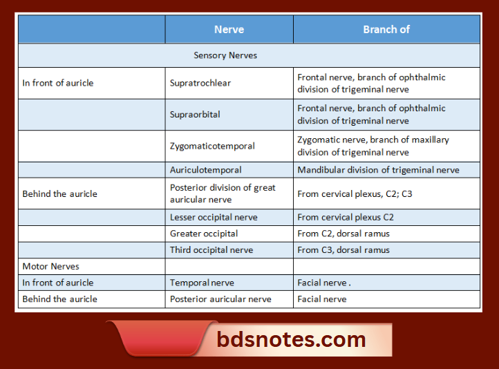
Sensory And Motor Nerve Supply Of The Scalp
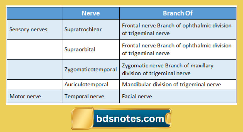
1. Sensory nerve supply
- In front of the auricle: branches are
- Supratrochlear nerve
- It is a branch of the frontal nerve which in turn is a branch of the ophthalmic division of a trigeminal nerve
- Supraorbital Nerve
- Branch of the frontal nerve which in turn is a branch of the ophthalmic division of a trigeminal nerve
- Zygomaticotemporal nerve
- Branch of the zygomatic nerve which is a branch of the maxillary division of a trigeminal nerve
- Auriculotemporal nerve
- It is a branch of the mandibular division of a trigeminal nerve
- Supratrochlear nerve
- Behind the auricle
- Posterior division of great auricular nerve
- Branch from cervical plexus C2, C3
- Lesser occipital nerve
- Branch from cervical plexus C2
- Greater occipital nerve
- Branch from C2, dorsal ramus
- Third occipital nerve
- Branch from C3, dorsal ramus
- Posterior division of great auricular nerve
“Factors influencing success with layers of the scalp studies: Q&A”
2. Motor nerves
- In front of the auricle
- Temporal nerve
- Branch of the facial nerve
- Temporal nerve
- Behind the auricle
- Posterior auricular
- Branch of the facial nerve
- Posterior auricular
Thus Scalp has its sensory nerve supply from branches of the trigeminal nerve and cervical plexus while the motor nerve supply is from the facial nerve
Layers of scalp BDS question
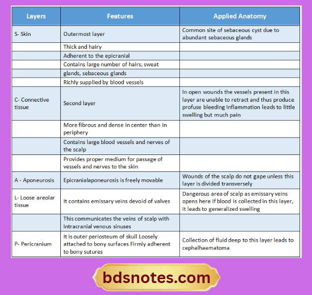
Loose Areolar Tissue Of Scalp And Its Applied Anatomy
4th layer of the scalp is loose areolar tissue
- Loose areolar tissue contains emissary veins devoid of valves
- This communicates the veins of the scalp with intracranial venous sinuses
Loose Areolar Tissue Extent:
- Anteriorly eyelids
- Posteriorly Highest and superior nuchal lines
- Each side has superior temporal lines
Loose Areolar Tissue Applied Anatomy:
- Loose Areolar Tissue is referred as a dangerous area of the scalp
- This is because of the opening of emissary veins here
- If blood is collected in this layer, it may lead to generalized swelling
Levator Palpebrae Superioris
- Levator Palpebrae Superioris is dilator of palpebral fissure
- Levator Palpebrae Superioris has a radial arrangement
- They are better developed around the eyes
Levator Palpebrae Superioris Origin:
- The inferior surface of the lesser wing of the sphenoid
- The orbital surface of the body of the sphenoid anterior to the optic foramen
“Steps to explain functions of the layers of the scalp: Protection vs thermoregulation: Q&A guide”
Levator Palpebrae Superioris Insertion:
- Orbital septum and palpebral ligaments
- Superior tarsal plate
- The skin of upper eyelid
Levator Palpebrae Superioris Nerve supply:
- Upper-division of an oculomotor nerve
- The sympathetic chain from the T1 segment of the spinal cord
Levator Palpebrae Superioris Applied anatomy:
- Paralysis of this muscle causes drooping of the upper eyelid called ptosis
Layers Of The Scalp With Its Applied Aspects
Layers of the scalp:
“Role of protection in shielding underlying structures: Questions answered”
Facial Nerve In The Face
- The facial nerve leaves the skull by passing through the stylomastoid foramen
- Next, it crosses the lateral side of the base of the styloid process
- Enters the posteromedial surface of the parotid gland
- Crosses the retromandibular vein and external carotid artery
- Behind the neck, it divides into five terminal branches which emerge along the anterior border of the parotid gland
Facial nerve in face Branches:
- Within the facial canal
- Greater petrosal nerve
- Nerve to the stapedius
- Chorda tympani nerve
- At its exit from stylomastoid foramen
- Posterior auricular
- Digastric
- Stylohyoid
- Terminal branches
- Temporal
- Zygomatic
- Buccal
- Marginal mandibular
- Cervical
- Communicating branches with adjacent cranial and spinal nerves
Retromandibular Vein
Retromandibular vein Formation:
- The retromandibular vein is formed by the union of the superficial temporal and maxillary vein
Retromandibular vein Course:
- Descends in the substance of parotid gland superficial to the external carotid artery
- Runs between ramus of the mandible and the sternocleidomastoid muscle
Retromandibular vein Branches:
- Anterior branch
- Joins anterior facial vein to form common facial vein
- Drains into the internal jugular vein
- Posterior branch
- Joins posterior auricular vein to form external jugular vein
Retromandibular vein Applied Anatomy:
- Parrot’s sign
- A retromandibular vein is the sensation of pain when pressure is applied to the retromandibular vein
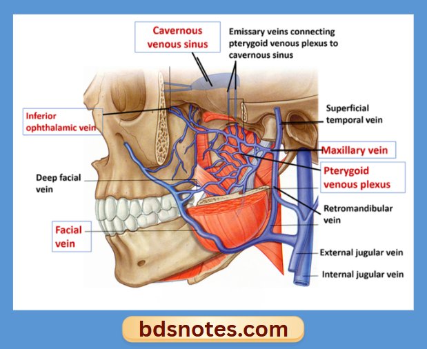
Venous Drainage Of The Face
Venous Drainage:
“Differential applications of pharmacological vs non-pharmacological treatments: Questions answered”
Veins of the face communicate with the cavernous sinus.
Occipitofrontalis Muscle
Occipitofrontalis muscles have two bellies
- Occipital or occipitalis
- Frontal or frontalis
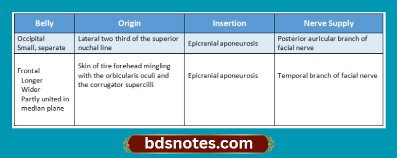
Actions:
- Raises the eyebrows
- Causes horizontal wrinkles in the skin of the forehead.
Question 20. Bell’s palsy
Answer:
Idiopathic paralysis of the facial nerve of sudden onset
Etiology 5 HYPOTHESIS:
- Rheumatic
- Cold
- Ischaemia
- Immunological
- Viral
Bell’s palsy Clinical Features:
- Pain in post auricular region
- Sudden onset
- Unilateral loss of function
- Loss of facial expression
- Absence of wrinkles on the forehead
- Loss of wrinkles
- Crocodile tears
- Inability to close the eye an effort to do so causes round or eyeball upwards
- Watering of eye
- Inability to blow the cheek
- Nasolabial fold disappears
- The tip of the nose is deviated
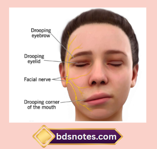
- Loss of taste sensation
- Hyperacusis
- Slurring of speech
Deep Facial Vein
- Deep connection of the facial vein includes communication between the supraorbital & pterygoid plexus through a deep facial vein, which passes backward over buccinators
- The facial vein communicates with the cavernous sinus through these connections
Deep facial vein Applied Anatomy:
- Infections from the face especially from the upper lip & the lower part of the face can spread in a retrograde direction & cause thrombosis of cavernous sinus
- This area is, therefore, called the dangerous area of the face
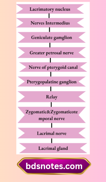
Secretomotor Supply To Lacrimal Gland.
“Can early intervention reverse layers of the scalp risks? FAQs provided”
Palpebral Ligament
- The palpebral fascia of the two lids forms the orbital septum
- Its thickening from tarsal plates of tarsi in the lids & palpebral ligaments at the angles
Dangerous Area Of The Scalp.
Loose areolar tissue
- Loose areolar tissue is the fourth layer of the scalp
- Loose areolar tissue is known as a “Dangerous area of the scalp” as an emissary vein opens here
- If blood is collected in this layer, it leads to generalized swelling
Question 25. Palatine aponeurosis
Answer:
The posterior border of the hard palate provides attachment to the palatine aponeurosis
Levator Palpebrae Superioris
- Levator palpebrae superioris is dilator of palpebral fissure
- Levator palpebrae superioris has radial arrangement
- They are better developed around the eyes
Levator palpebrae superioris Origin:
- The inferior surface of the lesser wing of the sphenoid
- The orbital surface of the body of the sphenoid anterior to the optic foramen
Levator palpebrae superioris Insertion:
- Orbital septum & palpebral ligaments
- Superior tarsal plate
- Skin of the upper eyelid
Levator palpebrae superioris Nerve Supply:
- Upper-division of the oculomotor nerve
- The sympathetic chain from the T1 segment of the spinal cord
Levator palpebrae superioris Applied Anatomy:
- Paralysis of this muscle causes drooping of the upper eyelid called ptosis
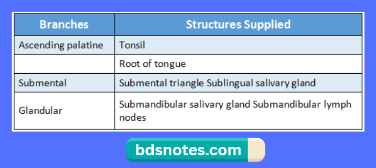
Cervical Branches Of The Facial Artery
- Ascending palatine nerve
- Tonsillar nerve
- Glandular nerve Submental nerve
Formation & Termination Of Anterior Facial Vein
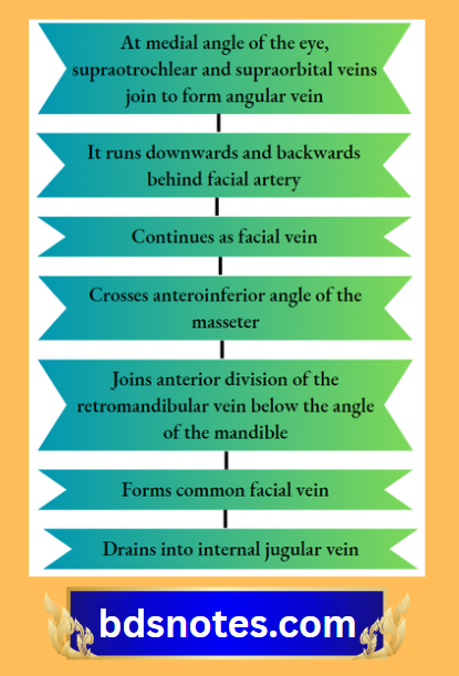
- Supratrochlear & supraorbital veins unite at the medial angle of the eye
- It forms an angular vein
- This continues and forms facial vein
- Then, facial vein along with the anterior division of the retromandibular vein forms a common facial vein
- This drains & terminates as an internal jugular vein
Facial Vein
- The facial vein is the largest vein of the face with no valves
Facial vein Origin:
- At a medial angle of the eye as an angular vein
Facial vein Course:
- The angular vein is formed by union of the supraorbital & supratrochlear vein
- The facial vein runs downward & backward behind the facial artery
- The facial vein crosses the anteroinferior angle of the masseter, pierces deep fascia, crosses the submandibular gland
- The facial vein joins the anterior division of the retromandibular vein below the angle of the mandible to form a common facial vein
Facial vein Terminates:
- The facial vein drains into the internal jugular vein.
“Asymptomatic vs symptomatic effects of delayed treatment: Answered”
Branches Of Facial Artery In The Face
1. Anterior branches
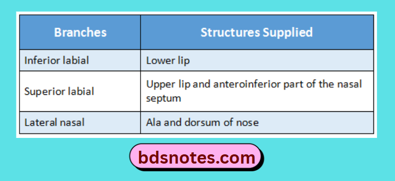
2. Posterior branches
- They are small and unnamed
Branches Of Facial Artery In The Neck
Retromandibular Vein
Retromandibular vein Formation:
- Retromandibular vein is formed by the union of the superficial temporal and maxillary vein
Retromandibular vein Course:
- Descends substance of parotid gland superficial to external carotid artery
- Runs between the ramus of mandible and the sternocleidomastoid muscle
Retromandibular vein Branches:
- Anterior branch
- Joins anterior facial vein to form common facial vein
- Drains into the internal jugular vein
- Posterior branch
- Joins posterior auricular vein to form external jugular vein
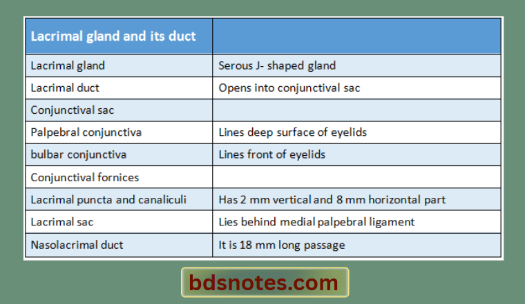
Nasolacrimal Duct
- Nasolacrimal duct is 18 mm long membranous passage
- The valve of Hasner, a fold of mucous membrane forming an imperfect valve is present at the lower end of the nasolacrimal duct
Nasolacrimal duct Course:
- Begins at lower end of the lacrimal sac
- Runs downwards, backward and laterally
- Opens into inferior meatus of nose
Applied anatomy:
Epiphora a condition where there is excessive secretion of lacrimal fluid overflowing on the cheeks may occur due to the obstruction in the lacrimal fluid pathway at the level of nasolacrimal duct
Galea Aponeurotica
- Galea aponeurotica is third layer of scalp
- Galea aponeurotica is freely movable
Galea aponeurotica Attachments:
- Anteriorlyinsertion of frontalis
- Posteriorlyinsertion of occipitalis
- In between occipital belliesexternal occipital protuberance and highest nuchal lines
- Each sideattached to superior temporal line
Galea aponeurotica Applied anatomy:
- Wounds of the scalp donot gape unless this layer is divided transversely
“Early warning signs of undiagnosed function-related issues: Common questions”
Structures Constituting Lacrimal Apparatus
- Lacrimal gland and its dud
- Conjunctival sac
- Lacrimal puncta and canaliculi
- Lacrimal snc
- Nasolacrimal duct
Dangerous Area Of Face
- The facial vein communicates with the cavernous sinus through its deep connections
- The facial vein is devoid of valves and rests directly on the facial muscles
- The movements of facial muscles facilitates the spread of emboli from the infected area of upper lip and lower part of the nose in retrograde direction and causes thrombosis of cavernous sinus
- Hence the area between the upper lip and lower part of nose is consider as dangerous area of face
Nerve Supply Of Scalp
“How does thermoregulation depend on scalp layers? FAQ explained”
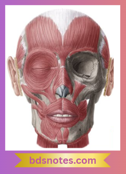
Name Any Six Muscles Of Facial Expressions.
Cutaneous Innervation Of Anterior Quadrant Of Scalp.
Lacrimal Gland
- Lacrimal gland is serous J shaped structure
Lacrimal gland Site:
- In the lacrimal fossa on the anterolateral part of the roof of the bony orbit and partly on the upper eyelid.
Lacrimal gland Parts:
- Orbital part larger and deeper
- Palpebral part smaller and superficial
Leave a Reply