Kesling Diagnostic Setup
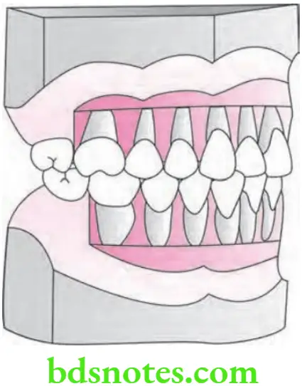
Kesling’s diagnostic setup.
The diagnostic setup was first proposed by HD Kesling in 1956.
Kesling’s Diagnostic Indication helps the clinician to estimate arch length discrepancy.
Kesling’s Diagnostic Procedure
- Both the maxillary and mandibular studying casts of the patient are prepared.
- Trimming of the base of both the study casts is done parallel to the occlusal plane.
- In the mandibular cast, horizontal cuts are made 3 mm apical to the gingival margin with the help of a fret saw blade.
- Vertical cuts are also made to separate the individual teeth.
- Trimming of mesial and distal ends of the root is done to facilitate the seating at the new position.
- In the formed slit during the cutting of the teeth, wax blocks are placed.
“Benefits Of Using Kesling Diagnostic Setup”
- On the study cast, the mandibular incisor teeth are arranged at an angle of 65° to Frankfort’s horizontal plane.
- Canines, as well as premolars, should be placed in correct contact relationships.
- If the remaining space is inadequate for receiving fist molars, extraction should be indicated. First premolars should be eliminated from the setup and placed with second premolars in contact with canines.
- Cutting of maxillary teeth is carried out, and they are repositioned in a wax set-up by articulating them with a mandibular set-up.
“Risk Factors For Errors In Kesling Diagnostic Setup”
Kesling’s Diagnostic Indication
- Kesling’s Diagnostic Indication is useful in visualizing and testing the effect of complex tooth movement and extractions on the occlusion.
- Patients can be motivated by simulating the various corrective procedures on the cast.
- Tooth size arch length discrepancy can be visualized using a set-up.
- Kesling’s Diagnostic Indication acts as a guide for extractions in the treatment plan.
Leave a Reply