Inflammatory Cells
Describe inflammatory cells.
Answer:
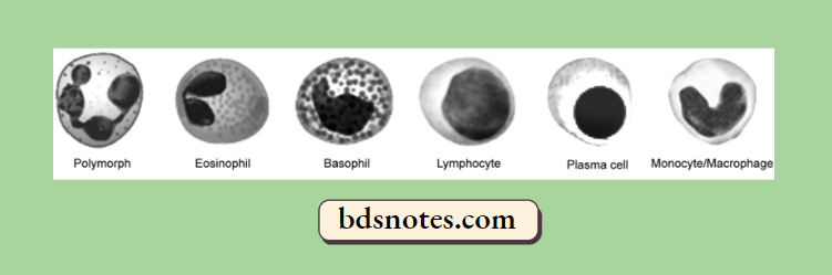
Following are the inflammatory cells i.e.
Circulating leucocytes, i.e. polymorphonuclear neutrophils, eosinophils, basophils, and lymphocytes
Plasma cells
Tissue macrophages.
Inflammatory cells
Inflammatory cells Polymorphonuclear Neutrophils
- Commonly called neutrophils or polymorphs, these cells along with basophils and eosinophils are known as granulocytes due to the presence of granules in the cytoplasm.
- These granules contain many substances like proteases, myeloperoxidase, lysozyme, esterase, aryl sulfatase, acid and alkaline phosphatase, and cationic proteins.
- The diameter of neutrophils ranges from 10 to 15 µm and is actively motile.
- These cells comprise 40-75% of circulating leucocytes and their number is increased in blood (neutrophilia) and tissues in acute bacterial infections.
- These cells arise in the bone marrow from stem cells.
- The functions of neutrophils in inflammation are as follows:
- Initial phagocytosis of micro-organisms as they form the first line of body defense in bacterial infection. The steps involved are the adhesion of neutrophils to vascular endothelium, emigration through the vessel wall, chemotaxis, engulfment, degranulation, killing, and degradation of the foreign material.
- Engulfment of antigen-antibody complexes and nonmicrobial material.
- The harmful effects of neutrophils are the destruction of the basement membranes of glomeruli and small blood vessels.
inflammatory cells Eosinophils
- These are larger than neutrophils but are fewer in number, comprising 1 to 6% of total blood leucocytes.
- Eosinophils share many structural and functional similarities with neutrophils like their production in the bone marrow, locomotion, phagocytosis, lobed nucleus, and presence of granules in the cytoplasm containing a variety of enzymes, of which major basic protein and eosinophil cationic protein is the most important which have bactericidal and toxic action against helminthic parasites. However, granules of eosinophils are richer in myeloperoxidase than neutrophils and lack lysozyme.
- High level of steroid hormones leads to a fall in the number of eosinophils and even disappearance from blood.
- The absolute number of eosinophils is increased in the following conditions and, thus, they take part in inflammatory responses associated with these conditions:
- Allergic conditions
- Parasitic infestations
- Skin diseases
- Certain malignant lymphomas.
Role of neutrophils in inflammation
Inflammatory cells Basophils
- Basophils comprise about l% of circulating leucocytes and are morphologically and pharmacologically similar to mast cells of the tissue.
- These cells contain coarse basophilic granules in the cytoplasm and a polymorphonuclear nucleus.
- These granules are laden with heparin and histamine.
- Basophils and mast cells have receptors for IgE and degranulate when cross-linked with antigens.
- The role of these cells in inflammation are:
- In the immediate and delayed types of hypersensitivity reactions
- Release of histamine by IgE-sensitised basophils.
Inflammatory cells Lymphocytes
- These cells are the most numerous of the circulating leucocytes (20-45%).
- Apart from blood, lymphocytes are present in large numbers in the spleen, thymus, lymph nodes, and mucosa-associated lymphoid tissue (MALT).
- They have scanty cytoplasm and consist almost entirely of a nucleus.
- Besides their role in antibody formation (B lymphocytes) and in cell-mediated immunity (T lymphocytes), these cells participate in the following types of inflammatory responses:
- In tissues, they are dominant cells in chronic inflammation and late-stage acute inflammation.
- In blood, their number is increased in chronic infections like tuberculosis.
Inflammatory cells Plasma Cells
- These cells are larger than lymphocytes with more abundant cytoplasm and an eccentric nucleus that has a cartwheel pattern of chromatin.
- Plasma cells are normally not seen in peripheral blood.
- They develop from lymphocytes and are rich in RNA and ?-globulin in their cytoplasm. There is an inter-relationship between plasmacytosis and hyperglobulinemia. These cells are most active in antibody synthesis.
- Their number is increased in the following conditions:
- Prolonged infection with immunological responses, For Example. in syphilis, rheumatoid arthritis, tuberculosis
- Hypersensitivity reactions
- Multiple myeloma.
Mononuclear-Phagocyte System
Following is the role of macrophages in inflammation:
- Phagocytosis (cell eating) and pinocytosis (cell drinking).
- Macrophages on activation by lymphokines released by T lymphocytes or by non-immunologic stimuli elaborate a variety of biologically active substances such as:
- Proteases like collagenase and elastase degrade collagen and elastic tissue.
- Plasminogen activator which activates the fibrinolytic system.
- Products of complement.
- Some coagulation factors convert fibrinogen to fibrin.
- Chemotactic agents for other leucocytes.
- Metabolites of arachidonic acid.
- Growth-promoting factors for fibroblasts, blood vessels, and granulocytes.
- Cytokines like interleukin-1 and tumor necrosis factor.
- Oxygen-derived free radicals.
Leave a Reply