Hemangioma
Hemangiomas are lesions which are not present at birth but they manifest within the fist month of life, exhibit a rapid proliferative phase and slowly involute to nonexistent.
Classification of Hemangiomas by Watson and McCarthy
- Capillary hemangioma
- Cavernous hemangioma
- Angioblastic hemangioma
- Racemose hemangioma
- Diffse systemic hemangioma
- Metastaizing hemangioma
- Port-Wine stain
- Hereditary hemorrhagic telangiectasia
“Early Signs Of Hemangioma In Infants”
hemangioma
Hemangiomas are lesions which are not present at birth but they manifest within the fist month of life, exhibit a rapid proliferative phase and slowly involute to nonexistent.
Classification of Hemangiomas by Watson and McCarthy
- Capillary hemangioma
- Cavernous hemangioma
- Angioblastic hemangioma
- Racemose hemangioma
- Diffse systemic hemangioma
- Metastaizing hemangioma
- Port-Wine stain
- Hereditary hemorrhagic telangiectasia
“Difference Between Hemangioma And Other Skin Growths”
Hemangioma Clinical Features
- Occur most commonly in infants and children.
- Peak incidence of central hemangiomas is during 2nd decade of life.
- More common in females
- Most commonly affected bones are facial bones, i.e. mandible, maxilla and nasal bones.
- Lesion appears as a flat or raised lesion of mucosa which is deep red or bluish red and is circumscribed.
- Lesion is compressible and filed slowly when released.
- Intra-orally commonly affected sites are lip, tongue, buccal mucosa and palate.
“Understanding The Role Of Blood Vessels In Hemangioma”
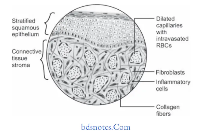
Hemangioma Histopathology
- There are several histopathologic types of hemangioma found in oral cavity, among them two very common types are:
- Capillary hemangioma
- Cavernous hemangioma.
Capillary hemangioma
- They are histologically characterized by numerous, small,endothelial lined capillaries in lesion which are densely packed by erythrocytes.
- Cells of endothelial lining are single layered.
- Endothelial cells are spindle shaped.
- Capillaries are well formed and are present throughout the lesion.
- Fibrous connective tissue stroma is not well formed and is loosely arranged.
infantile hemangioma
“The Role Of Imaging In Diagnosing Hemangioma Accurately”
“Step-By-Step Guide To Diagnosing Hemangioma Effectively”
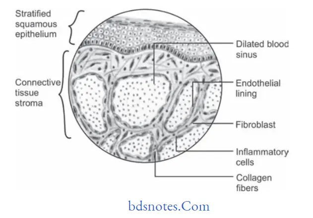
Histopathology Of Cavernous Hemangioma
- They are histologically characterized by large, irregularly shaped, dilated, endothelialized sinuses which contain large aggregates of erythrocytes.
- A single layer of flttd endothelial cell lines each sinus.
- Sinus lacks muscular coat on their walls.
- Large area of hemorrhage and hemosiderin pigments is often seen within cavernous hemangioma lesions.
“Comprehensive Overview Of Hemangioma And Its Types”
Leave a Reply