Gingivectomy
Answer. Gingivectomy means excision of the gingiva.
By removing the pocket wall-gingivectomy provides visibility and accessibility for complete calculus removal and thorough smoothening of the roots, creating a favorable environment for gingival healing and restoration of a physiologic gingival contour.
“Risk Factors For Complications After Gingivectomy”
“Purpose Of Gingivectomy In Dentistry”
Surgical Gingivectomy
The step-by-step technique for gingivectomy is as follows:
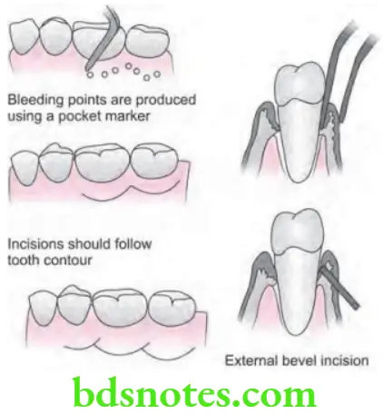
- Step 1: Periodontal pocket should be mapped out over the external gingival surface by inserting a probe to the bottom of pocket and puncturing external surface of gingiva at the depth of probe penetration.
Gingivectomy Procedure Explained
- Step 2: Periodontal knives i.e. Kirkland knives are indicated for incisions on the facial and lingual surfaces. Orban periodontal knives are indicated for inter-dental incisions. Bard–Parker (BP) blades (#12 and #15), and scissors are indicated as an auxiliary instruments.
“Early Signs Of Problems During Gingivectomy Recovery”
- External bevel incision should be started apical to the points marking course of pockets, and it is directed coronally to a point between base of the pocket and crest of the bone. This should be close to the bone without exposing it to remove soft tissue coronal to the bone.
- Exposure of bone is undesirable, if this occurs, healing presents minimal complications if the area is adequately covered by the surgical dressing. Either interrupted or continuous incisions can be used.
- Incision should be beveled at approximately 45 degrees to tooth surface, and it should re-create the normal festooned pattern of gingiva. Failure to bevel the incision will leave a broad, fibrous plateau which will delay development of physiologic contour.
Gingivectomy Healing Process
- Step 3: Eliminate the excised pocket wall, irrigate the area and examine root surface.
- Step 4: Undergo Scaling and root planing.
- Step 5: Cover the area by surgical dressing.
“Best Ways To Understand Gingivectomy Surgery”
Healing Following Gingivectomy
- Healing after surgical gingivectomy is basically by secondary intention:
- The initial response is the formation of a productive surface clot.
- The clot is then replaced by granulation tissue.
- Within 24 hours, there is an increase in new connective tissue cells mainly angioblasts and by third day numerous fibroblasts are located in this area.
- The highly vascular granulation tissues grow coronally, creating a new free gingival margin and sulcus.
Gingivectomy Postoperative Care
- Capillary derived from blood vessels of periodontal ligament migrate into the granulation tissue, and with in two weeks they connect with gingival vessels.
“Importance Of Gingivectomy For Gum Health”
- After 12 to 24 hours, epithelial cells at the margins of the wound start to migrate over the granulation tissue, separating it from the contaminated surface layer of clot. Epithelial activity at the margins reaches to maximum in 24 to 36 hours.
- The new epithelial cells arise from the basal and deeper spinous layers of the wound, edge epithelium and migrate over the wound over a fibrin layer that is later resorbed and replaced by connective tissue bed.
- The epithelial cells advance by tumbling action, with the cells becoming fixed to the substrate by hemidesmosomes and a new basement lamina.
Gingivectomy Indications And Techniques
- After 5 to 14 days, surface epithelialization is generally complete. During the first 4 weeks after gingivectomy, keratinization is less than it was before surgery. Complete epithelial repair take about 1 month.
- Vasodilation and vascularity begin to decrease after the fourth day of healing and appear to be almost normal by 16th day.
- Complete repair of the connective tissue take about 7 weeks.
Leave a Reply