Gingiva Definition
“What are the microscopic and macroscopic features of the gingiva?”
Gingiva is the part of the oral mucosa that covers the alveolar process of the jaws and surrounds the neck of the teeth.
Gingiva is masticatory mucosa.
Gingiva Macroscopic Features
Marginal Gingiva (Unattached Gingiva)
- Marginal Gingiva is the terminal edge or border of the gingiva surrounding the teeth in collar-like fashion.
- Marginal Gingiva is usually 1 mm wide.
- Marginal Gingiva forms the soft tissue wall of the gingival sulcus.
Gingival Sulcus
- The gingival sulcus is the shallow crevice or space around the tooth bonded by the surface of the tooth on one side and marginal gingiva and the epithelium, lining the free margin of the gingiva on other side.
- Gingival Sulcus is ‘V’ shaped.
- Under ideal conditions, the depth of the gingival sulcus is about ‘0’.
- Clinically, healthy gingiva in human, the normal gingival sulcus depth is 2 to 3 mm.
“Understanding the role of gingival anatomy in oral health”
Read And Learn More: Periodontics Question And Answers
Attached Gingiva
- Attached gingiva is the part of the gingiva which is tightly attached to the underlying periosteum of alveolar bone and cementum by connective tissue fibers.
- Attached gingiva is fim, resilient and hence immovable portion of the gingiva.
- Attached gingiva is thus, firmly entrenched between two movable structures: the marginal gingiva coronally and the alveolar mucosa apically.
- Attached gingiva is different from keratinized gingiva and should not be confused with it.
“How do microscopic and macroscopic features differ in the gingiva?”
- Width of the attached gingiva is measured as the distance between mucogingival junction and the projection on the external surface of the bottm of gingival sulcus/periodontal pocket. The dimensions of the attached gingiva vary from the anterior to the posterior teeth.
- Width of the attached gingiva is generally wider in the maxilla than in the mandible. The narrowest zone of the gingiva is found in the region of the maxillary and mandibular 1st premolars and usually in connection with frenum and muscle attachments. The pattern of variation is approximately the same in deciduous and permanent teeth.
- Width of the attached gingiva is wider in the supraerupted teeth. It also increases with age. This increase in dimension occurs as a result of an increase in the height of the alveolar process, which in turn, is the result of passive eruption.
- Width of attached gingiva is greater in incisor region that is 3.5 to 4.5 mm in maxilla and 3.3 to 3.9 mm in mandible and is less in posterior segments that is there is least width in premolar region which is 1.9 mm in maxilla and 1.8 mm in mandible.
“Importance of studying gingival features for dental professionals”
Interdental Gingiva/Papilla
- Part of the gingiva which fils the inter-dental space between two adjacent teeth is known as inter-dental papilla.
- It appears pyramidal or triangular from the facial and lingual aspect along with its lateral borders. Tip of the pyramid is formed by continuation of marginal gingiva of adjacent teeth.
- In the posterior region, interdental gingiva has a ‘tent’ shape.
Gingival Col
- Facial as well as lingual portions of interdental papillae forms the concave or valley like area which fits below the contact area. The valley like area is known as Col.
- Col or Gingival col is lined by non-keratinized epithelium.
- Gingival Colconfirms the shape of interproximal contact areas.
- Because of the non-keratinized epithelium, col is prone to inflammation and lead to disease.
- In this area oral hygiene accessibility is not possible.
“Common challenges in identifying gingival features under a microscope”
Gingival Microscopic Features
- Gingiva consists of an area of connective tissue covered by stratified squamous epithelium.
- Gingival Epithelium is divided into three types i.e. outer epithelium, sulcular epithelium and junctional epithelium.
Gingival Outer/Oral Epithelium
- Oral epithelium, also called as an outer epithelium, is a keratinized stratifid squamous type of epithelium.
- It covers the crest and outer surface of marginal gingiva and surface of attached gingiva.
- Oral epithelium consists of following types of cellular layers:
- Stratum basale: Cells of stratum basale are either cylindrical or cuboid. Basal cells are found immediately next to the connective tissue and are separated from connective tissue by a basement membrane. It is the germinative layer and hence can divide. When two daughter cells have been formed by cell division, an adjacent older basal cell is pushed into the spinous cell layer and starts as a keratinocyte to traverse the epithelium. It takes approximately 1 month for a keratinocyte to reach the outer epithelial surface, where it is shed from the stratum corneum.
“Steps to teach students about gingival microscopic and macroscopic features”
- Stratum spinosum: It is a prickle cell layer in which large polyhedral cells with short cytoplasmic processes are present. The uppermost cells from this layer contain granules called as keratinosomes or Odland bodies. These are modified lysosomes, which contain a large amount of enzymes acid phosphatase that is involved in the destruction of organelle membranes.
- Stratum granulosum: Cells of this layer are flattned in plane parallel to the gingival surface. Keratohyalin granules, which are associated with keratin formation, are round in shape and appear within the cytoplasm of the cell.
- Stratum corneum: It consists of closely packed, flattened cells that have lost nuclei and the most other organelles as they become keratinized. The cells are densely packed with tonofiaments. Clear, rounded bodies probably representing lipid droplets appear within the cytoplasm of the cell.
Sulcular Epithelium
- Sulcular Epithelium lines the gingival sulcus.
- Sulcular Epithelium is a thin, non-keratinized stratifid squamous epithelium without rete pegs.
- Sulcular Epithelium extends from coronal limit of the junctional epithelium to the crest of the gingival margin.
- Sulcular Epithelium usually shows many cells with hydropic degeneration.
- Sulcular Epithelium is devoid of granulosum and corneum strata.
- The sulcular epithelium has the potential to keratinize, if
- It is reflected and exposed to oral cavity.
- The bacterial flora of the sulcus is totally eliminated.
- Sulcular Epithelium is extremely important because it may act as a semipermeable membrane through which infectious bacterial products pass into the gingiva and tissue fluid from the gingiva seeps into the sulcus.
“Role of diagrams and models in explaining gingival anatomy”
Junctional Epithelium
- Junctional epithelium consists of a collar-like band of stratified squamous non-keratinized epithelium.
- Junctional Epithelium is 3-4 layer thick in early life but increases with age upto 10 to 20 layer.
- The length of junctional epithelium ranges from 0.25 to 1.35 mm.
- These cells can be grouped in two strata: the basal layer that faces the connective tissue and the suprabasal layer that extends to the tooth surface.
- The proliferative layer which leads to most of the cell divisions is located in contact with the connective tissue. Shedding surface of junctional epithelium is located at the coronal end which forms bottm of gingival sulcus.
- Junctional epithelium is formed by conflence of the oral epithelium and the reduced enamel epithelium at the time of eruption.
- Junctional epithelial cells are interconnected by few desmosomes and occasionally by gap junctions.
- Junctional epithelium, mainly at its basal cell layers is innervated by the sensory nerve fibers.
“How do textbooks explain gingival histology and macroscopic features?”
- Division of junctional epithelium is there in three zones i.e. coronal, middle and apical.
- Junctional epithelium is attached to the tooth surface by means of an internal basal lamina and to the gingival connective tissue by an external basal lamina which has the same structure as other epithelial–connective tissue attachments elsewhere in the body.
- External basal lamina consists of same structure and the composition as other basement membranes, elsewhere in the body while the internal basal lamina has different structural and molecular characteristics. This lacks the common basement membrane components i.e. collagen Type 4 and 7, most laminins forms.
- The internal basal lamina consists of a lamina densa (adjacent to the enamel) and a lamina lucida to which hemidesmosomes are attached. Hemidesmosomes have a decisive role in the fim attachment of the cells to the internal basal lamina on the tooth surface.
- Junctional epithelial cells migrate in the coronal direction to free surface where they desquamate. As the surface area occupied by basal cells is more than bottm of sulcus, exfoliation occurs at extremely high rate.
- Attachment of the junctional epithelium to the tooth is reinforced by the gingival fibers, which brace the marginal gingiva against the tooth surface. So, the junctional epithelium and gingival fibers are considered as functional unit. Dentogingival unit: Dentogingival unit = junctional epithelium + gingival fibers.
- Intercellular spaces of junctional epithelium give a pathway for fluid and transmigrating the leukocytes. In absence of clinical signs of inflammation, approximately 30,000 PMNs migrate per minute via junctional epithelium of all human teeth in the oral cavity.
Principal Cells of the Gingival Epithelium
- Keratinocytes
- Non-keratinocytes
- Langerhans cells
- Merkel cells
- Melanocytes
The main function of the gingival epithelium, is to protect the deep structure by proliferation and differentiation of the keratinocytes.
Keratinocytes
- They are the principal cells of gingival epithelium.
- Keratinocytes form the keratinized layer outer epithelium by proliferation and differentiation.
- Proliferation of keratinocytes take place by mitosis in basal layer these proliferated cells begin to migrate to the surface.
- Differentiation involves the process of keratinization, which consists of a sequence of biochemical and morphologic events that occur in the cell as it migrates from basal layer.
- Main morphologic change: Flattning of cells with long tonofiaments. Prevention of keratohyaline granule and disappearance of nucleus.
- Complete keratinization process leads to formation of orthokeratin.
- In parakeratin layer the stratum cornea retains pyknotic nuclei and the keratohyaline granules are dispersed.
- The uppermost cells of the stratum spinosum contains dense granules, i.e. keratinosome or Odland bodies which are modified lysosome.
Non-keratinocytes
- Melanocytes: These are dendritic cells located in the basal and spinous layers of the gingival epithelium.
- They synthesize melanin organelles called premelanosome/melanosome.
- Melanin are found or phagocytosed within the melanophages or melanophors.
- Langerhans cells: Dendritic cells located among the keratinocytes at all suprabasal level.
- These are modified monocytes derived from bone marrow.
- They have important role in immune reaction.
- They contain g-specific granules (Birbeck’s granule).
- They found in oral epithelium of normal gingiva, less in sulcular epithelium and absent in junctional epithelium.
- Merkel cells:
- Located in deeper layer of epithelium.
- Harbor the nerve endings and are connected to the adjacent epithelium by desmosome.
- They contain tactile receptors.
Gingival Fluid (Sulcular Fluid)
- Gingival fluid can be represented as either a transudate or an exudate.
- Gingival fluid consists of vast array of biochemical factors, thereby offring its potential use as a diagnostic or prognostic biomarker of the biologic state of the periodontium in health and disease.
- It also consists of components of connective tissue, epithelium, inflmmatory cells, serum, and microbial flora that inhabit the gingival margin or the sulcus.
- In healthy sulcus, the amount of gingival fluid is very small.
- During inflammation, the gingival fluid flow increases, and its composition starts to resemble that of an inflammatory exudate.
- The main route of the gingival fluid diffusion is via the basement membrane, through the relatively wide intercellular spaces of the junctional epithelium, and then into the sulcus.
- The gingival fluid is believed to do the following:
- Cleanse material from the sulcus
- Gingival fluid contain plasma proteins that may improve adhesion of the epithelium to the tooth
- Gingival fluid possess antimicrobial properties
- Gingival fluid exerts antibody activity to defend the gingiva.
“Can interactive tools improve learning outcomes?”
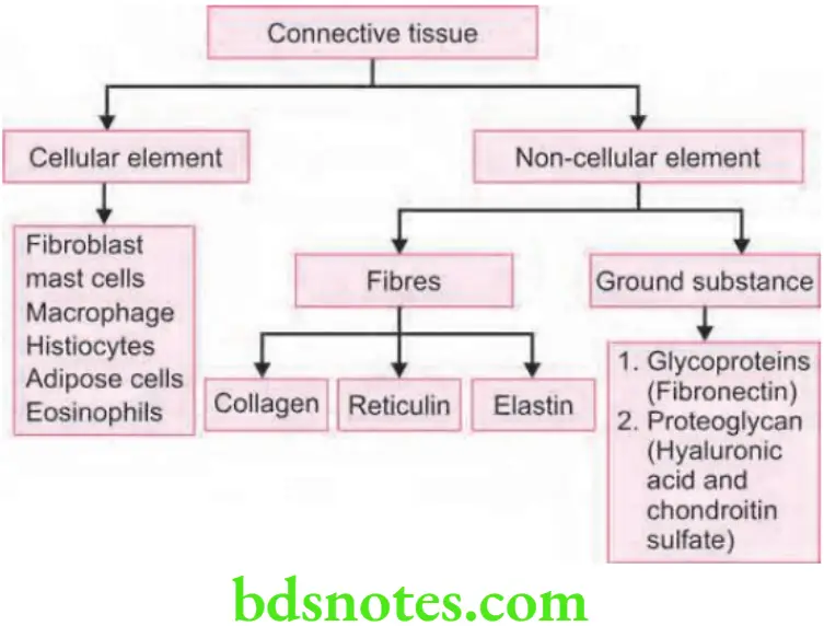
Gingival Connective Tissue
- The major components of the gingival connective tissue are collagen fibers i.e. about 60% by volume, fibroblasts i.e. 5%, vessels, nerves, and matrix i.e. about 35%.
- The connective tissue of the gingiva is called as lamina propria and it consists of two layers i.e.
- A papillary layer subjacent to the epithelium that consists of papillary projections between the epithelial rete pegs
- A reticular layer that is contiguous with the periosteum of the alveolar bone.
- Connective tissue has a cellular compartment and an extracellular compartment composed of fibers and ground substance.
- The ground substance fils the space between fibers and cells; it is amorphous, and it has a high water content. It is composed of proteoglycans (mainly hyaluronic acid and chondroitin sulfate) and glycoproteins (mainly fironectin).
- The three types of connective tissue fibers are collagen, reticular and elastic.
- Collagen type 1 forms the bulk of the lamina propria and provides the tensile strength to the gingival tissue. Type 4 collagen branches between the collagen type 1 bundles, and it is continuous with fibers of the basement membrane and the blood vessel walls.
Gingival Fibers
The gingival fibers are arranged in three groups:
Gingivodental
- The gingivodental fibers are those on the facial, lingual and interproximal surfaces.
- They are embedded in the cementum just beneath the epithelium at the base of gingival sulcus.
“Asymptomatic vs symptomatic effects of poor educational resources”
- On the facial and lingual surfaces, they project from cementum in fan-like confirmation towards the crest of alveolar ridge and the outer surface of marginal gingiva.
- They terminate on periosteum of bone or in the attached gingiva.
Circular Group
Circular group of fiber are course through connective tissue of the marginal and interdental gingiva and encircle the tooth in ring like fashion.
Transseptal Group
- They are located interproximally.
- It is formed by horizontal bundles that extend between the cementum of approximating teeth into which they are embedded.
- They lie in the area between epithelium at base of gingival sulcus and crest of interdental bone.
“Early warning signs of knowledge gaps in gingival education”
Other
Semicircular Fibers
Semicircular fibers are attached to the proximal surface of tooth just below the CEJ and go around facial or lingual marginal gingiva of the tooth, and attach on the other proximal surface of same tooth.
Transgingival Fibers
Attach in the proximal surface of one tooth, transverse interdental space and go around the facial/lingual surface of adjacent tooth, again traverse the interdental space and attach in proximal surface of next tooth.
Leave a Reply