Giant Cells
Giant cells are multinucleated cells that commonly occur at sites of chronic inflammation mostly complicated by granuloma formation.
Besides giant cells occurring in various lesions, there also exists physiological giant cells by virtue of the presence of multinucleated nature,present as a part of healthy tissues.
Multinucleated giant cells are highly stimulated cells of the monocytemacrophage lineage at a terminal stage of maturation and studies have demonstrated that their multinucleated appearance is produced by cell–cell fusion rather than by nuclear division without cytokinesis.
giant cells
“Risk Factors For Diseases Involving Giant Cells”
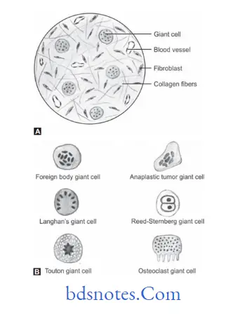
Classification of Giant Cells
Giant cell can be classified based according to their occurrence in the body:
- Physiological giant cells:
- Osteoclast
- Striated muscle cells
- Megakaryocytes
- Syncytiotrophoblast
“Early Signs Of Giant Cell Involvement In Diseases”
- Pathological giant cells:
- Langhan’s giant cells
- Foreign body giant cells
- Touton giant cells
- Tumor giant cells
- Warthin–fikeldey giant cells
Osteoclast
Osteoclasts are highly specialized for resorption of bone mineral and matrix through the coordinated secretion of hydrogen ions and proteolytic enzymes.
Although most osteoclasts are large multinucleated cells, there are reports of mononuclear osteoclasts.
Situated on the bone surface, it occupies a concavity surface of small bone spicules.
An enlarged surface area created by plasma membrane infoldings, the ruffled border, characterizes the secretory or apical surface directed toward the bone.
“Difference Between Langhans And Foreign Body Giant Cells”
In routine histologic sections, the ruffl border appears striated and lightly stained. The presence of a ruffl border is an indication that the osteoclast is actively engaged in bone resorption.
Each ruffl border is surrounded by a clear zone (or sealing zone), a cytoplasmic area rich in cytoplasmic actin fiaments and devoid of major cytoplasmic organelles.
Through close adaptation of the cell surface to the bone matrix, the clear zone establishes a seal between the bone resorption compartment and the interstitial flid.
types of giant cells
“Role Of Macrophages In Forming Giant Cells”
Striated Muscle
They are the muscles in which the cells exhibit cross striations at the light microscopic level.
It is further sub-classifid based on its location:
- Skeletal muscle
- Visceral striated muscle
- Cardiac muscle
Skeletal Muscle Cell
A skeletal muscle cell is a multinucleated syncytium. In skeletal muscle, each muscle fier is a multinucleated syncytium.
A muscle fier is formed during development by the fusion of small, individual muscle cells called myoblasts.
“Understanding The Role Of Macrophages In Giant Cell Formation”
When viewed in cross section, the mature multinucleated muscle fiers reveal a polygonal shape with a diameter of 10 to 100 µm.
Visceral Striated Muscle
It is morphologically identical to skeletal muscle but is restricted to the soft tissues, namely the tongue, pharynx, lumbar part of diaphragm and upper part of esophagus.
These play an essential role in speech, breathing and swallowing.
“Step-By-Step Guide To Diagnosing Giant Cells Effectively”
Cardiac Muscle
Cardiac muscle has the same type and arrangement of the contractile fiament as skeletal muscle.
In addition, cardiac muscle fiers exhibit densely staining cross bands called intercalated disks, that cross the fiers in a linear fashion or frequently in a way that resembles the rosters of a stairway.
The intercalated disks represent a highly specialized attchment sites between adjacent cells.
The linear cell—cell attchment of the cardiac muscle cells results in ‘fiers’ of variable length.
Megakaryocytes
Platelets are derived from large polypoid cells (cells whose nuclei contain multiple sets of chromosomes) in bone marrow are called megakaryocytes.
Syncytiotrophoblast
- The syncytiotrophoblast is a continuous, normally uninterrupted layer that extends over the surfaces of all villous trees as well as over pAns of the inner surfaces chorionic and basal plates.
- The syncytiotrophoblast is a multinucleated protoplasmic mass without intercellular boundaries.
- From this mass emerge figer like projections, which penetrate through the endometrial epithelium into the endometrial stroma.
“Tips To Prevent Complications From Giant Cell-Related Diseases”
Langhans giant Cell
They are characterized by location of the nuclei at the periphery of the cell in an acute confiuration.
They are seen in lesions such as tuberculosis, sarcoidosis, leprosy and vasculitis.
These are special, more highly organized forms than are ‘foreign body’ multinucleated giant cells.
These giant cells may attin diameters of 40 to 50 µm. They have a large mass of cytoplasm containing 20 or more small nuclei arranged peripherally.
Foreign body giant Cell
Formation of foreign body giant cell is hallmark of the foreign body reaction and is harmful to implanted biomaterials because it contributes to the degradation of the biomaterial and leads to stress cracking, tissue firosis and a chronic response.
Foreign body giant cells are thought to be a source of chemokines that mediate the neutrophils and lymphocytes.
“Comprehensive Overview Of Giant Cells And Their Significance”
Touton Giant Cell
Touton giant cells are characterized by a ring of nuclei surrounding central eosinophilic zone and surrounded by a zone of pallor extending to the periphery of cell.
These giant cells are seen in lesions with high lipid content such as xanthoma, xanthogranuloma, fat necrosis.
The characteristic appearance of ‘Xanthelasmatic giant cell’ of Touton is determined merely by the presence of demonstrable lipid in the cytoplasm.
Tumor Giant Cell
A feature of anaplasia is the formation of tumor giant cells,some possessing only a single huge polymorphic nucleus and others having two or more nuclei.
These giant cells are not to be confused with inflammatory LanghAns or foreign body giant cells, which are derived from macrophages and contain many small normalappearing nuclei.
In the cancer giant cell, the nuclei are hyperchromatic and large in relation to the cell.
“Best Practices For Treating Giant Cell-Related Symptoms Safely”
Warthin-Finkeldey Giant Cell
The Warthin Finkeldey cells have up to 100 nuclei and contain spherical eosinophilic intracytoplasmic and intranuclear inclusions.
They are present in viral infections like measles.
Tompkins and Macaulay reported Warthin-Finkeldey giant cells in nasal secretions before the appearance of other clinical signs of measles such as Koplik’spots and skin rash.
These cells are found throughout the reticuloendothelial system and contain up to 100 nuclei.
“The Role Of Histopathology In Identifying Giant Cells Accurately”
Leave a Reply