Gastrointestinal Diseases Important Question And Answers
Question 1. Write Short notes on Stomatitis.
Or
Write Notes On Stomatitis.
Answer. Stomatitis is the inflammation of the mouth and is caused by bacterial, viral, and fungal infections in persons with poor oral hygiene or with blood dyscrasias.
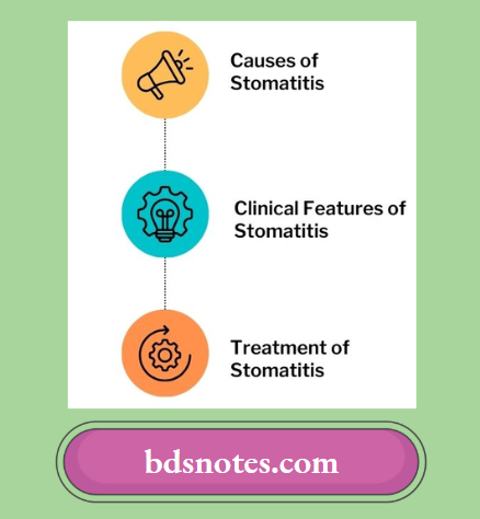
Causes of Stomatitis
- Local Causes of Stomatitis
- Poor oral hygiene
- Excessive use of tobacco
- Alcohol and spices
- Use of broad-spectrum antibiotics
- Drugs such as iodine or gold.
“Understanding gastrointestinal diseases through FAQs: Causes, symptoms, and treatments explained”
- General Causes of Stomatitis
The main general causes are infectious diseases. There are various types of infective stomatitis:- Bacterial, e.g. streptococcal stomatitis and Vincent’s stomatitis
- Viral, e.g. herpes simplex and herpes zoster
- Fungal, e.g. candidiasis and actinomycosis
- Recurrent aphthous stomatitis
- Mucocutaneous diseases, e.g. Lichen planus, pemphigus vulgaris, lupus erythematous, etc.
- Miscellaneous, e.g. diabetes, uremia, and drug toxicity.
“Importance of studying gastrointestinal diseases for healthcare professionals: Questions explained”
“Common challenges in diagnosing and treating gastrointestinal diseases effectively: FAQs provided”
Read And Learn More: General Medicine Question And Answers
Clinical Features of Stomatitis
- Lip, tongue, and gums are inflamed, swollen, and painful.
- The tongue is furred and a foul smell is present.
- Sometimes ulceration of the mucus membrane is present when a person is suffering from infectious stomatitis.
- The patient feels pain and difficulty in opening the mouth.
- There was an *excoriation and redness of the mucus membrane of the oral cavity.
“Steps to explain gastrointestinal diseases: Causes vs symptoms vs treatment: Q&A guide”

“Factors influencing success with gastrointestinal disease knowledge: Q&A”
General Medicine BDS 3rd Year Question and Answers
Treatment of Stomatitis
- If the drug is the causative factor discontinuation of the drug is done.
- It allergen is the causative factor remove allergen.
- Antihistaminic drugs such as cetirizine are to be given to the patient.
- Topical corticosteroid application should be done.
- Triamcinolone acetate is effective.
- Tetracycline mouthwash should be given which should be used four times a day for 5 to 7 days.
- Nutritional supplements are to be given such as vitamin B12, iron, and folic acid.
Common Gastrointestinal Disorders: Symptoms, Diagnosis, and Treatment
Leave a Reply