Ewing’s Sarcoma
Ewing’s sarcoma is a small round cell tumor. It arises inside the bone.
Ewing’s Sarcoma Clinical Features
- It occurs predominantly in children and young adults between 5 to 25 years with median age of 13 years.
- It is more common in males as compared to females.
- Whites develop this tumor more commonly but in blacks not even single case is reported.
- Pain in Ewing’s sarcoma is intermittnt in nature.
- Swelling of the bone is earliest clinical sign.
- Long bones of extremities are more commonly affcted besides these skull, clavicle, ribs, shoulder are also affcted.
ewing’s sarcoma
“Risk Factors For Developing Ewing’S Sarcoma”
- Ewing’s sarcoma in occur more commonly in mandible as compared to maxilla
- Paresthesia and loosening of teeth are common fidings in Ewing’s sarcoma of jaw.
- Appearance of jaw swelling is rapid and intraoral mass become ulcerated.
- Tumor commonly penetrates the cortex resulting in soft tissue mass which overlie affected area of bone.
Ewing’s Sarcoma Radiographic Features
- Most common fiding is formation of layers of new subperiosteal bone which produces onion skin appearance of fim.
- There is presence of irregular lytic bone destruction with ill-defied margins.
“Early Signs Of Ewing’S Sarcoma In Children And Teens”
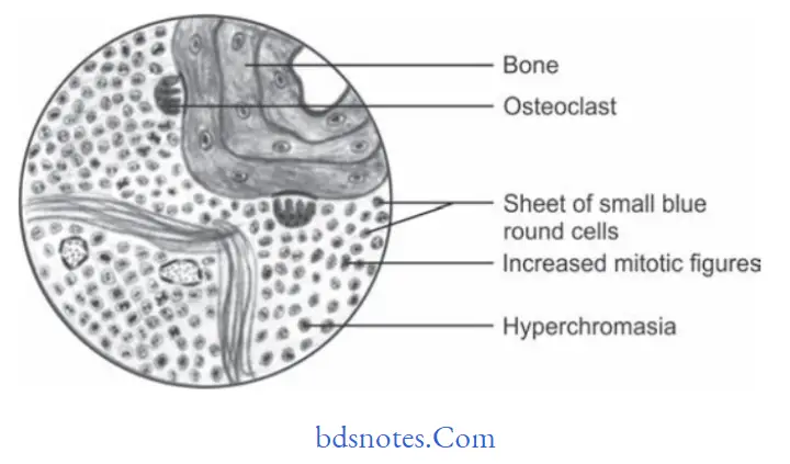
Ewing’s Sarcoma Histopathology
- Ewing’s sarcoma is highly cellular which consists of solid sheets or mass of small round cells with litte stroma, few connective tissue septa can also be seen.
- Cells are small and round in shape having scanty cytoplasm. Nuclei of cell is large round to oval in shape with dispersed chromatin and hyperchromasia.
- Borders of cell are indistinct.
- Cells are arranged in filigree pattern.
ewing sarcoma treatment
“Role Of Genetics In Causing Ewing’S Sarcoma”
- Mitotic figures are commonly seen.
- Multiple small vascular channels are also present.
- Hemorrhage with vascular lakes or sinuses can be seen.
- There is also presence of geographic necrosis with perivascular sparing.
- Necrosis can be seen on the opposite side of fragment of bone.
Ewing’s Sarcoma Histological Differential Diagnosis
- Small cell osteosarcoma
- Peripheral neuroectodermal tumor of infancy
- Metastatic neuroblastoma
- Mesenchymal chondrosarcoma
- Malignant lymphoma
- Embryonal rhabdomyosarcoma.
“Understanding The Role Of Genetics In Ewing’S Sarcoma”
Ewing’s Sarcoma Treatment
- Radical surgical excision should be done alone or coupled with Xray radiation.
- Current treatment consists of combined surgery, radiotherapy and multidrug chemotherapy which led to 40 to 80% of survival rates.
Leave a Reply