Anatomy And Physiology Of The Ear
Write a short note on ear ossicles.
Answer.
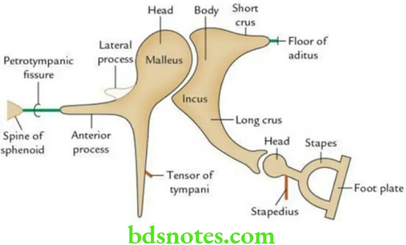
- There are three tiny bones present in the middle ear cavity.
- From medial to lateral, these are malleus (hammer), incus (anvil), and stapes (stirrup).
Malleus is the lateral ossicle. It has a head, neck, and three processes, i.e. handle, lateral process, and anterior process.
- The head articulates with the incus
- The handle passes downwards and is attached to the tympanic membrane. The medial aspect of the handle receives insertion of tensor tympani.
- The anterior process is attached to the spine of the sphenoid by a ligament.
- The lateral process is attached to the tympanic sulcus (bony) by anterior and posterior malleolar folds.
“Importance Of The Ear In Hearing And Balance”
Incus is the middle bone. It has a body, a short process, and a long process.
- The body articulates with the head of the malleus.
- A short crus is attached to the floor of aditus by a ligament.
- Long crus articulates with the head of stapes.
Stapes is the most medial (innermost) ossicle. It has a head, neck, anterior and posterior limbs, and footplate.
- The head articulates with the long process of the incus.
- Footplate is held in fenestra vestibule (oval window) by annular ligament.
- The neck of the stapes receives insertion of the stapedius.
“Risk Factors For Ear-Related Disorders Like Hearing Loss”
“Early Signs Of Issues With Ear Anatomy Or Function”
Ear Ossicles Applied anatomy
The vibrations of sound waves are transmitted from the tympanic membrane to the fluid (perilymph) of the inner ear by the ossicular chain.
- Paralysis of stapedius leads to hyperacusis.
- Otosclerosis (abnormal ossification of the annular ligament that anchors the stapes footplate leads to conduction deafness).
Inner Ear: Anatomy, Function & Related Disorders
Briefly describe the internal ear.
Answer.
- The internal ear is involved in both hearing and balance.
- It consists of two components: a membranous labyrinth and a bony (osseous) labyrinth.
“Understanding The Role Of The Auditory Nerve In Hearing”
Internal Ear Membranous labyrinth
It consists of four membranous parts/structures:
- Cochlear duct
- Saccule
- Utricle
- Three semicircular ducts
All these parts are interconnected to each other to form a labyrinth.
Internal Ear Functions
- The sensory receptor within the cochlear duct is a spiral organ of the Corti. It is concerned with hearing.
- The sensory receptors present within the saccule and utricle are maculae. They are concerned with static balance.
- The sensory receptors within the semicircular ducts are cristae ampullaris. They are concerned with kinetic balance.
“Best Practices For Diagnosing And Treating Ear Disorders”
Internal Ear Bony labyrinth It consists of intercommunicating bony spaces in the petrous part of the temporal bone.
The bony labyrinth consists of three parts:
- Cochlea
- Vestibule
- Three semicircular canals
Anatomy And Physiology Of The Ear
Write a short note on ear ossicles.
Answer.
- There are three tiny bones present in the middle ear cavity.
- From medial to lateral, these are malleus (hammer), incus (anvil), and stapes (stirrup).
Malleus is the lateral ossicle. It has a head, neck, and three processes, i.e. handle, lateral process, and anterior process.
- The head articulates with the incus
- The handle passes downwards and is attached to the tympanic membrane. The medial aspect of the handle receives insertion of tensor tympani.
- The anterior process is attached to the spine of the sphenoid by a ligament.
- The lateral process is attached to the tympanic sulcus (bony) by anterior and posterior malleolar folds.
Incus is the middle bone. It has a body, a short process, and a long process.
- The body articulates with the head of the malleus.
- A short crus is attached to the floor of aditus by a ligament.
- Long crus articulates with the head of stapes.
“The Role Of The Vestibular System In Maintaining Balance”
Stapes is the most medial (innermost) ossicle. It has a head, neck, anterior and posterior limbs, and footplate.
- The head articulates with the long process of the incus.
- Footplate is held in fenestra vestibule (oval window) by annular ligament.
- The neck of the stapes receives insertion of the stapedius.
Ear Ossicles Applied anatomy
The vibrations of sound waves are transmitted from the tympanic membrane to the fluid (perilymph) of the inner ear by the ossicular chain.
- Paralysis of stapedius leads to hyperacusis.
- Otosclerosis (abnormal ossification of the annular ligament that anchors the stapes footplate leads to conduction deafness).
Leave a Reply