Development of the Face, Lip, and Palate: A Step-by-Step Guide
Development Of The Face
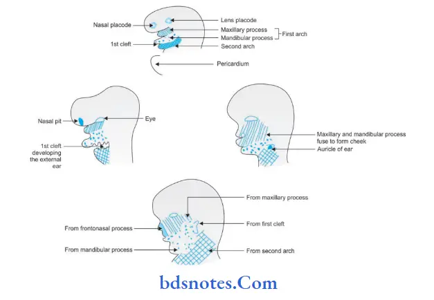
The face is derived from the following structures that lie around the stomatodeum, i.e.
- Frontonasal process
- The first pharyngeal (or mandibular) arch of each side.
- At this stage, each mandibular arch forms the lateral wall of the stomatodaeum.
- This arch gives off a bud from its dorsal end. This bud is called the maxillary process.
It grows ventromedial cranial to the main part of the arch which is now called the mandibular process.
“Importance Of Facial Prominence Fusion In Development”
- The ectoderm overlying the frontonasal process soon shows bilateral localized thickenings, that are situated a little above the stomatodeum. These are called the nasal placodes.
The formation of these placodes is induced by the underlying forebrain. The placodes soon sink below the surface to form nasal pits. - The pits are continuous with the stomatodeum below.
The edges of each pit are raised above the surface.
The medial raised edge is called the medial nasal process and the lateral edge is called the lateral nasal process.
“Best Ways To Understand Lip And Palate Development”
Development Of Lower Lip
Mandibular processes of the two sides grow towards each other and fuse in the midline.
They now form the lower margin of the stomatodeum.
If it is remembered that the mouth develops from the stomatodeum, it will be readily understood that the fused mandibular processes give rise to the lower lip and to the lower jaw.
“Risk Factors For Abnormal Face And Palate Development”
Development Of Upper Lip
Each maxillary process now grows medially and fuses, first with the lateral nasal process and then with the medial nasal process.
The median and lateral nasal processes also fuse. In this way, the nasal pits are cut off from the stomatodeum.
The maxillary processes undergo considerable growth.
At the same time, the frontonasal process becomes much narrower from side to side, with the result that the two external nares come close together.
The stomatodeum is now bounded above by the upper lip which is derived as follows:
“Early Signs Of Issues With Facial And Palate Formation”
- The mesodermal basis ofthe lateral part ofthe lip is formed from the maxillary process. The overlying skin is derived from the ectoderm covering this process.
- The mesodermal basis of the median part of the lip (called philtrum) is formed from the frontonasal process.
The ectoderm ofthe maxillary process, however, overgrows this mesoderm to meet that of the opposite maxillary process in the midline. As a result, the skin of the entire upper lip is innervated by maxillary nerves.
The muscles of the face are derived from the mesoderm of the second branchial arch and are therefore supplied by the facial nerve.
Development Of Nose
The nose receives contributions from the frontonasal process and the medial and lateral nasal processes of the right and left sides.
External nares are formed when the nasal pits are cut off from the stomatodeum by the fusion of the maxillary process with the medial nasal process.
External nares gradually approach each other.
“Role Of Neural Crest Cells In Face And Palate Formation”
Mesoderm becomes heaped up in the median plane to form the prominence of the nose.
‘Simultaneously, a groove appears between the regions of the nose and the bulging forebrain.
As the nose becomes prominent the external nares come to open downwards.
The external form of the nose is thus established.
Development Of Cheeks
After the formation of the upper and lower lips, the stomatodeum (which can now be called the mouth) is very broad.
In its lateral part, it is bounded above by the maxillary process and below by the mandibular process.
These processes undergo progressive fusion with each other to form the cheeks.
Development Of Eye
The region of the eye is first seen as an ectodermal thickening, the lens placode, which appears on the ventrolateral side of the developing forebrain, lateral and cranial to the nasal placode.
The lens placode sinks below the surface and is eventually cut- off from the surface ectoderm.
The developing eyeball produces a bulging in this situation.
The bulging of the eyes is at first directed laterally and lies in the angles between the maxillary processes and the lateral nasal processes.
With the narrowing of the frontonasal process, they come to face forward.
The eyelids are derived from folds of ectoderm formed above and below the eyes, and by mesoderm enclosed within the folds.
“Understanding The Role Of Neural Crest Cells In Facial Development”
Development Of External Ear
The external ear is formed around the dorsal part of the first ectodermal cleft.
A series of mesodermal thickenings (often called tubercles or hillocks) appear on the mandibular and hyoid arches where they adjoin this cleft.
The pinna (or auricle) is formed by the fusion of these thickenings.
Development Of Nasal Cavities
Nasal cavities are formed by extension of the nasal pits. These pits are at first in open communication with the stomatodeum.
Soon the medial and lateral nasal processes fuse and form a partition between the pit and the stomatodeum.
This is called the primitive palate and is derived from the frontonasal process.
The nasal pits now deepen to form the nasal sacs which expand both dorsally and caudally.
“Step-By-Step Guide To Explaining Facial And Palate Development”
The dorsal part of this sac is, at first, separated from the stomatodeum by a thin membrane called the bucconasal membrane (or nasal fi). This soon breaks down.
The nasal sac now has a ventral orifice that opens on the face (anterior or external nares) and a dorsal orifice that opens into the stomatodeum (primitive posterior nasal aperture).
The two nasal sacs are at first widely separated from one another by the frontonasal process.
However, the frontonasal process becomes progressively narrower.
“Tips To Prevent Abnormalities In Facial And Palate Development”
This narrowing of the frontonasal process, and the enlargement of the nasal cavities themselves, bring them closer together.
This intervening tissue becomes much thinned to form the nasal septum.
The ventral part of the nasal septum is attached below to the primitive palate.
More posteriorly the septum is at first attached to the bucconasal membrane, but on disappearance of this membrane it has a free lower edge.
The nasal cavities are separated from the mouth by the development of the palate.
The lateral wall of the nose is derived, on each side, from the lateral nasal process.
The nasal conchae appear as elevations on the lateral wall of each nasal cavity.
The original olfactory placodes form the olfactory epithelium that lies in the roof, and adjoining parts of thin walls, of the nasal cavity.
“The Role Of Prenatal Care In Ensuring Healthy Facial And Palate Formation”
Congenital abnormalities of lip and Palate
Following are the congenital abnormalities oflip and palate:
- Congenital lip pits
- Commissural lip pits
- Double lip
- Cleft lip and cleft palate.
Congenital lip Pits
Congenital lip is also known as a congenital fistula.
Congenital lip Pathogenesis
Congenital lip occurs due to the failure of the union of the embryonic sulcus of the lip which leads to persistent lateral sulci on the embryonic mandibular arch.
Congenital Lip Clinical Features
- It more commonly occurs in females.
- The vermilion border of the lip is commonly involved. The lower lip is involved.
- Lips appear swollen
- The lesion is present in the form of depression.
- On palpation, mucous secretion is seen from the base of the lip pit.
Congenital lip Treatment
Surgical excision is done.
Commissural lip Pits
They are mucosal invagination which arises at the vermilion border of the lip.
Commissural lip Pathogenesis
Its occurrence is due to the failure of normal fusion of embryonic maxillary and mandibular processes.
“How To Live A Healthier Life By Supporting Proper Facial Development”
Commissural Lip Clinical Features
- Males are commonly affected
- It presents as a unilateral or bilateral pit at the corners of the mouth on the vermilion border
- Its size ranges from a shallow depression to an open tract measuring 4 mm
- On palpation, less amount of saliva oozes out.
Commissural Lip Treatment
Surgical excision is done.
Double Lip
Double Lip is a fold of excessive tissue over the inner mucosa of the lip.
Double Lip Pathogenesis
It arises during the second week of gestation because of the persistence of the sulcus between pars glabrosa and pars villosa of the lip.
“Comprehensive Overview Of Facial And Palate Development Stages”
Double Lip Clinical Features
- The inner aspect of the lip is involved.
- At times when the upper lip becomes tensed, the double lip gives the appearance of a cupid bow.
Double Lip Treatment
Surgical excision is done.
Leave a Reply