Dentigerous Cysts: Causes, Symptoms, And Treatment
It is also known as follicular odontoma.
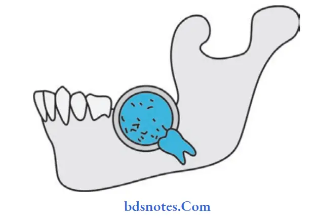
It is a common odontogenic cyst of epithelial origin, which encloses the crown of an impacted tooth at its neck portion.
The cyst is lined by squamous epithelium, surrounded by connective tissue.
Within the cyst, the tooth lies obliquely or sometimes embedded in the wall of the cyst.
As cyst grows, it displace the teeth deeper and deeper and prevent from eruption.
Dentigerous Cyst Surgery
“Step-By-Step Guide To Diagnosing A Dentigerous Cyst”

Read and learn More Cysts: Types, Causes, Symptoms, and Treatment
Dentigerous Cyst Diagnosis
Dentigerous Cyst Clinical Features
- Age and sex: It is usually found in children, equal in both the sex.
- Site: Most common site is mandibular third molar and maxillary canine which are most commonly impacted.
- Symptoms: Cyst remains asymptomatic, if uninfected. On infection inflammatory signs are present.
- Expansion of mandible: Since the inner table of mandible is strong the expansion mainly occurs in the outer aspect of the mandible.
The bone gets thinned out resulting in egg shell cracking. - Blue-domed cyst: When it contains blood then it is called as blue-domed cyst.
- A dentigerous cyst has the potential to become an aggressive lesion with the expansion of bone and subsequent facial asymmetry.
- There is extreme displacement of teeth, severe root resorption of adjacent teeth and pain.
“Tips To Prevent Complications From A Dentigerous Cyst“
“Effective Ways To Manage Dentigerous Cyst Recovery”
Dentigerous Cyst Radiographic Features
- It is a well-defined radiolucency usually associated with hyperostotic borders unless it is secondarily infected.
- Bony margins are well-defied and sharp.
- It may involve the crown symmetrically; it may expand from the crown.
- Large cysts are confined to the mandible. There may be resorption of roots.
Dentigerous Cyst In Children
“The Role Of X-Rays In Diagnosing A Dentigerous Cyst“
Dentigerous Cyst Differential Diagnosis
- Ameloblastoma or ameloblastic firoma: They are multilocular and not associated with crown of an unerupted teeth.
- Adenomatoid odontogenic tumors: They are rare and occur in the maxillary anterior region.
- Calcifying odontogenic cyst: It occurs as peri coronal radiolucency and contains evidence of calcification.
- Developmental primordial and follicular primordial cyst: It occurs in the crown of the uner updated tooth and super position of the image which may cause cyst-like radiolucency to appear as a dentigerous cyst on the radiograph.
“Comprehensive Overview Of Dentigerous Cyst Symptoms“
Dentigerous Cyst Treatment
- Treatment via an intraoral approach or extraoral is decided by the size ofcyst, adequate access and whether it desirable to save the involved tooth.
- Marsupialization: It is indicated in children if the cyst is very large in the size and involved tooth/teeth are to be maintained.
The tooth may erupt in occlusion as the defect heals with normal bone or orthodontic forces may be used to bring the tooth into occlusion. - Enucleation: Alternatively cyst can be enucleated together with involved tooth in adults as possibility of tooth eruption is low.
Leave a Reply