Cranial Nerves
Question 1. Enumerate the cranial nerves.
Answer. Following are the cranial nerves:
- I Olfactory (Smell)
- II Optic (Sight)
- III Oculomotor (Moves eyelid and eyeball and adjusts the pupil and lens of the eye)
- IV Trochlear (Moves eyeballs)
- V Trigeminal (Facial muscles including chewing; Facial sensations)
- VI Abducens (Moves eyeballs)
- VII Facial (Taste, tears, saliva, facial expressions)
- VIII Vestibulocochlear (Auditory)
- IX Glossopharyngeal (Swallowing, saliva, taste)
- X Vagus (Control of PNS, e.g. smooth muscles of GI tract)
- XI Accessory (Moving head and shoulders, swallowing)
- XII Hypoglossal (Tongue muscles—speechand swallowing)
Cranial Nerves
“Optic Nerve Damage And Symptoms”
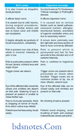
Question 2. Describe differentiating features of Bell’s palsy and trigeminal neuralgia.
Answer.
Read And Learn More: Neurological and Facial Disorders: Causes, Diagnosis, and Treatment Strategies
“Oculomotor Nerve Palsy Causes And Treatment”
Cranial nerves are a fascinating and essential part of our nervous system. They consist of twelve pairs of nerves that arise directly from the brain, each serving unique functions related to sensory and motor control. Understanding these nerves is crucial for anyone interested in neuroanatomy, as they play a vital role in our daily lives, affecting everything from our ability to see and smell to how we move our facial muscles. This guide aims to break down the complexities of cranial nerves, making it easier to grasp their significance and functions.
Key Takeaways
- Cranial nerves are twelve pairs of nerves that originate from the brain, each with distinct sensory or motor functions.
- They are classified into sensory, motor, or mixed nerves, impacting senses like smell, vision, and movement.
- Key cranial nerves include the olfactory nerve for smell, the optic nerve for vision, and the facial nerve for facial movements.
- Cranial nerves have specific pathways and exit points from the skull, connecting to various brain regions.
- Understanding cranial nerves is important for diagnosing and treating neurological disorders.
12 Cranial Nerves
Understanding Cranial Nerves
Definition and Overview
“Trochlear Nerve Injury And Recovery”
Cranial nerves are a set of twelve nerves that emerge directly from the brain, in contrast to spinal nerves, which emerge from the spinal cord. These nerves are responsible for a wide array of functions, primarily serving the structures of the head and neck. Each cranial nerve has a specific name and number (I-XII), which indicates its function and position, respectively. They play a vital role in sensory perception, motor control, and various autonomic functions.
Importance in Neuroanatomy
Understanding cranial nerves is really important in neuroanatomy because they connect the brain to different parts of the head, neck, and even some areas of the torso. They’re like the superhighways for signals that control everything from eye movement to swallowing. If there’s damage to a cranial nerve, it can cause very specific problems, which helps doctors figure out where the issue is in the brain. Knowing the cranial nerves anatomy helps in diagnosing and treating a bunch of neurological conditions.
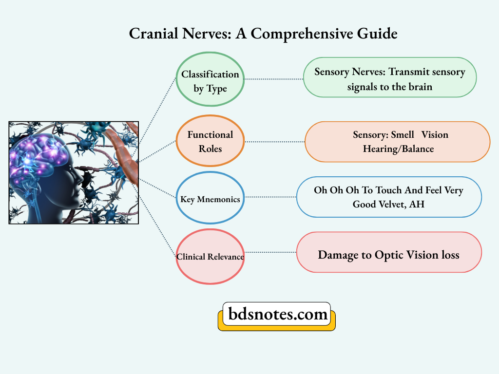
Classification of Cranial Nerves
Cranial nerves can be classified based on their primary function:
- Sensory Nerves: These nerves are primarily involved in transmitting sensory information from the body to the brain. Examples include the olfactory nerve (smell), optic nerve (vision), and vestibulocochlear nerve (hearing and balance).
- Motor Nerves: These nerves primarily control muscle movement. Examples include the oculomotor, trochlear, abducens, spinal accessory, and hypoglossal nerves.
- Mixed Nerves: These nerves have both sensory and motor functions. Examples include the trigeminal, facial, glossopharyngeal, and vagus nerves.
It’s also worth noting that some classify a fourth category: parasympathetic. These nerves carry parasympathetic fibers, which regulate involuntary functions like heart rate, digestion, and gland secretion. The oculomotor, facial, glossopharyngeal, and vagus nerves all contain parasympathetic fibers.
Here’s a quick summary:
“Common Disorders Of Cranial Nerves”
Sensory Functions of Cranial Nerves
Alright, let’s talk about the cranial nerves that are all about sensation. These guys are the reason you can smell your coffee in the morning, see the world around you, and hear your favorite tunes. It’s pretty amazing when you think about it.
Olfactory Nerve and Smell
So, the olfactory nerve (CN I) is the first one up, and it’s all about smell. Think of it as your personal scent detector. When you inhale, odor molecules hit the receptors in your nasal cavity, and boom, the olfactory nerve sends that info straight to your brain. Pretty simple, right? But without it, life would be pretty bland. Imagine not being able to smell cookies baking or the fresh scent after rain. That’s why detecting smells is so important!
Functions Of Cranial Nerves
Optic Nerve and Vision
Next up, we have the optic nerve (CN II), which is responsible for vision. This nerve transmits visual information from your retina to your brain. Light enters your eye, hits the retina, and the optic nerve takes it from there, creating the images you see. It’s like a high-speed data cable for your eyeballs. Problems with this nerve can lead to all sorts of vision issues, so keeping it healthy is key.
“Causes Of Cranial Nerve Damage”
Vestibulocochlear Nerve and Hearing
Last but not least, there’s the vestibulocochlear nerve (CN VIII). This one’s a double whammy because it handles both hearing and balance. The cochlear part deals with sound, while the vestibular part keeps you upright and oriented. Damage to this nerve can cause hearing loss, tinnitus (ringing in the ears), or balance problems like vertigo. Trust me, you don’t want to mess with your balance; it’s essential for everything from walking to riding a bike.
These sensory nerves are super important for how we experience the world. They take in all sorts of information and send it to the brain for processing. Without them, life would be a lot less colorful, noisy, and, well, smelly.
Here’s a quick rundown:
- Olfactory: Smell
- Optic: Vision
- Vestibulocochlear: Hearing and Balance
Motor Functions of Cranial Nerves
Oculomotor Nerve and Eye Movement
Okay, so the oculomotor nerve is a big deal when it comes to moving your eyes around. It’s basically the main controller for most of the muscles that handle eye movement. Think about it – looking up, down, and side to side? That’s largely the oculomotor nerve at work. It also takes care of lifting your eyelid and making sure your pupil does its thing, like getting smaller in bright light. If this nerve gets damaged, you might end up with a droopy eyelid, double vision, or trouble focusing. Not fun!
Facial Nerve and Facial Expressions
The facial nerve is what lets you smile, frown, and make all those other awesome facial expressions. It controls the muscles in your face, so you can show the world how you’re feeling. But it’s not just about expressions; it also handles some of your taste sensation and controls some glands, like the ones that make saliva and tears. Problems with the facial nerve can lead to facial paralysis, like in Bell’s palsy, where one side of your face droops. Imagine trying to eat or talk with that going on – it’s a real challenge. The facial nerve is super important for communication and basic functions.
Hypoglossal Nerve and Tongue Movement
The hypoglossal nerve is all about your tongue. It controls most of the muscles that let you stick it out, move it around, and, you know, talk and swallow. If this nerve isn’t working right, you might have trouble speaking clearly or swallowing food. It can also cause your tongue to weaken or twitch.
Think about how much we use our tongues every day without even realizing it. Talking, eating, even just keeping saliva where it belongs – the hypoglossal nerve is quietly working in the background to make it all happen. It’s easy to take it for granted until something goes wrong.
Here’s a quick rundown of what can happen if the hypoglossal nerve is damaged:
- Difficulty speaking (dysarthria)
- Trouble swallowing (dysphagia)
- Tongue weakness
- Tongue twitching (fasciculations)
Understanding how these nerves work helps to understand motor system organization.
“Symptoms Of Cranial Nerve Dysfunction“
Mixed Functions of Cranial Nerves
Some cranial nerves are real overachievers, handling both sensory and motor duties. It’s like they decided one job just wasn’t enough. These nerves are involved in everything from feeling sensations to controlling muscles, making them super important for everyday functions.
Trigeminal Nerve and Sensation
The trigeminal nerve (CN V) is a big deal. It’s responsible for sensation in your face, like when you feel a cool breeze or accidentally bite your tongue. But it also controls the muscles you use for chewing. So, if you’re enjoying a steak, thank your trigeminal nerve. Problems with this nerve can cause intense facial pain, like in trigeminal neuralgia. It’s divided into three main branches:
- Ophthalmic (V1): Sensation from the forehead, eyes, and nose.
- Maxillary (V2): Sensation from the cheeks, upper lip, and palate.
- Mandibular (V3): Sensation from the lower jaw and controls muscles of mastication.
Glossopharyngeal Nerve and Taste
The glossopharyngeal nerve (CN IX) is all about taste and swallowing. It handles taste sensations from the back of your tongue and controls some of the muscles in your throat that help you swallow. It also plays a role in salivation. Damage to this nerve can make it hard to taste things or swallow properly. It’s not something you think about until it’s not working right. The glossopharyngeal nerve also carries sensory information from the tonsils, pharynx, and middle ear.
Functions Of Cranial Nerves
Vagus Nerve and Autonomic Functions
The vagus nerve (CN X) is the longest cranial nerve and a true multitasker. It’s a major player in the autonomic nervous system, which controls things you don’t consciously think about, like heart rate, digestion, and breathing. It also has sensory and motor functions related to your throat and voice box. The vagus nerve influences a wide range of bodily functions. It’s like the body’s internal internet, connecting the brain to many different organs. Here’s a quick look at its diverse roles:
- Regulates heart rate and blood pressure.
- Stimulates digestive organs.
- Controls muscles in the throat for speech and swallowing.
“Treatment Options For Cranial Nerve Disorders”
The vagus nerve’s extensive reach means that problems with it can cause a variety of symptoms, from digestive issues to changes in heart rate. It’s a complex nerve with a big job.
Understanding the mixed functions of these cranial nerves is key to understanding how our bodies work. They show how sensation and movement are often intertwined, allowing us to interact with the world around us. The facial and trigeminal nuclei are complex structures that highlight this integration.
Anatomy of Cranial Nerves
Origin and Pathways
Okay, so when we talk about cranial nerves, we’re really talking about twelve special nerves that do a bunch of important jobs, mostly in your head and neck. These nerves are unique because, unlike most nerves that come from your spinal cord, these guys pop directly out of your brain. It’s kind of like having VIP access to the control center. Two of them, the olfactory and optic nerves, start in the forebrain. The accessory nerve actually has roots in the spinal cord, which is a bit of an oddball. The rest? They all come from the brainstem. Each nerve then takes its own special route to get where it needs to go, whether it’s to control a muscle, relay a sensation, or both.
Exit Points from the Skull
Ever wonder how these nerves actually get out of your skull? Well, the skull isn’t just one solid bone; it’s got all sorts of holes and openings called foramina. Each cranial nerve has its own designated exit point, like a secret passage. The order in which they exit, from front to back (rostral to caudal, if you want to get fancy), is actually how they got their Roman numeral names (I to XII). So, the olfactory nerve (CN I) exits the skull way up front, while the hypoglossal nerve (CN XII) makes its exit much further back. It’s like a carefully planned highway system for your nervous system.
Cranial Nerve Anatomy
Connections to the Brainstem
So, the brainstem is where most of these cranial nerves really get their start. Think of it as the central hub. The brainstem itself is made up of a few parts: the midbrain, the pons, and the medulla oblongata. Each cranial nerve has a specific nucleus (or group of nerve cells) within the brainstem that it’s connected to. These nuclei are like little command centers, and they’re responsible for processing the signals that come in and go out through the cranial nerves. It’s a pretty complex setup, but it’s what allows these nerves to do everything from controlling your eye movements to helping you taste your food.
Understanding the anatomy of the cranial nerves is super important because it helps us figure out what’s going on when things go wrong. If a nerve is damaged or compressed, knowing its pathway and where it exits the skull can help doctors pinpoint the problem and figure out the best way to fix it.
Here’s a quick rundown of the cranial nerves:
“Surgical Repair Of Cranial Nerve Damage”
- Olfactory (I): Smell
- Optic (II): Vision
- Oculomotor (III): Eye movement
- Trochlear (IV): Eye movement
- Trigeminal (V): Sensation in the face, chewing
- Abducens (VI): Eye movement
- Facial (VII): Facial expressions, taste
- Vestibulocochlear (VIII): Hearing and balance
- Glossopharyngeal (IX): Taste, swallowing
- Vagus (X): Autonomic functions, like heart rate and digestion
- Accessory (XI): Shoulder and neck movement
- Hypoglossal (XII): Tongue movement
Clinical Significance of Cranial Nerves
Cranial nerves are super important. When something goes wrong with them, it can tell doctors a lot about what’s happening in your brain. It’s like they’re little messengers, and if one of them is delivering the wrong message, it’s a sign something’s up.
Cranial Nerve Anatomy
Common Disorders and Symptoms
Okay, so what happens when cranial nerves go haywire? Well, it depends on the nerve. If it’s the optic nerve, you might have vision problems. If it’s the facial nerve, you could get Bell’s palsy, where one side of your face droops. Problems with the vestibulocochlear nerve can cause hearing loss or dizziness. The symptoms really vary depending on which nerve is affected.
Here’s a quick rundown:
- Vision changes
- Facial paralysis
- Hearing loss
- Balance issues
- Difficulty swallowing
“Medications For Managing Cranial Nerve Pain”
Diagnostic Tests for Cranial Nerve Function
So, how do doctors figure out if there’s a problem with your cranial nerves? They do a cranial nerve exam. It’s not as scary as it sounds. They’ll ask you to do things like follow a light with your eyes, smile, stick out your tongue, and shrug your shoulders. Each of these actions tests a different nerve. For example, testing cranial nerve anatomy is essential for almost any medical specialty.
Here are some common tests:
- Visual field testing
- Fundoscopy
- Facial muscle strength assessment
Cranial Nerve Overview
Treatment Approaches for Cranial Nerve Issues
Treatment really depends on what’s causing the problem. If it’s an infection, antibiotics might help. If it’s a tumor pressing on a nerve, surgery or radiation might be needed. Sometimes, it’s just a matter of managing the symptoms with medication or therapy. Early diagnosis is key for effective treatment.
Sometimes, the cause isn’t clear, and treatment focuses on relieving symptoms and improving quality of life. This might involve pain medication, physical therapy, or speech therapy, depending on the specific nerve affected and the resulting symptoms.
Cranial Nerve Examination Techniques
Neurological Assessment Basics
Okay, so before we jump into the specifics, let’s talk about the basics of a neurological exam. It’s not just about poking and prodding; it’s about observing. We’re looking at things like a person’s mental state, their posture, how they move, and their reflexes. These observations give us clues before we even start testing individual cranial nerves. It’s like gathering intel before the main mission. You want to make sure the patient is comfortable and can understand your instructions. A quiet room helps too, minimizing distractions. This sets the stage for accurate testing.
Specific Tests For Each Nerve
Alright, now for the fun part – testing each of the twelve cranial nerves. Each nerve has its own special test. For example, to check the optic nerve function (CN II), we test vision and visual fields. For the facial nerve (CN VII), we look for facial symmetry by having the person smile, frown, and raise their eyebrows. And so on. Here’s a quick rundown:
- Olfactory Nerve (CN I): Smell test (usually with coffee or peppermint).
- Optic Nerve (CN II): Visual acuity and visual fields.
- Oculomotor, Trochlear, Abducens Nerves (CN III, IV, VI): Eye movements and pupil response.
- Trigeminal Nerve (CN V): Facial sensation and jaw movement.
- Facial Nerve (CN VII): Facial expressions and taste (anterior tongue).
- Vestibulocochlear Nerve (CN VIII): Hearing and balance.
- **Glossopharyngeal Nerve (CN IX): Taste (posterior tongue) and swallowing.
- Vagus Nerve (CN X): Gag reflex and voice quality.
- Accessory Nerve (CN XI): Shoulder shrug and head rotation.
- Hypoglossal Nerve (CN XII): Tongue movement.
“Physical Therapy For Cranial Nerve Recovery”
Interpreting Examination Results
So, you’ve done all the tests. Now what? Interpreting the results is where the real skill comes in. You’re looking for asymmetries, weaknesses, or any deviation from the norm. A subtle weakness in one side of the face could indicate a facial nerve issue. An abnormal gag reflex might point to a problem with the vagus nerve. It’s like being a detective, piecing together the clues to figure out what’s going on. Remember to consider the patient’s medical history and other symptoms. It’s all part of the bigger picture.
It’s important to remember that cranial nerve exams are just one piece of the puzzle. They should always be interpreted in the context of a full neurological evaluation and the patient’s overall clinical presentation. Don’t jump to conclusions based on a single test result. Look at the whole picture.
Cranial Nerve Overview
Wrapping It Up
So, there you have it! We’ve taken a good look at the cranial nerves and what they do. These twelve nerves play a big role in how we interact with the world, from seeing and smelling to moving our face and neck. Each one has its own job, and together, they help keep everything running smoothly. Understanding them can seem tough at first, but breaking it down like this makes it a bit easier. Whether you’re a student or just curious, knowing about cranial nerves is pretty handy. They’re more than just a bunch of names; they’re key players in our daily lives.
Frequently Asked Questions
What Are Cranial Nerves?
Cranial nerves are twelve nerves that come directly from the brain. They help us with our senses and movements.
Why Are Cranial Nerves Important?
Cranial nerves are important because they control many functions in our head and neck, like seeing, hearing, and moving our face.
How Are Cranial Nerves Classified?
Cranial nerves can be classified into three types: sensory, motor, or mixed, which means they can do both functions.
“Case Studies On Cranial Nerve Outcomes”
What Is An Example Of A Sensory Cranial Nerve?
An example of a sensory cranial nerve is the optic nerve, which helps us see.
What Role Does The Facial Nerve Play?
The facial nerve is a motor cranial nerve that controls our facial expressions, like smiling or frowning.
How Can Doctors Check If Cranial Nerves Are Working Properly?
Doctors can perform specific tests to check each cranial nerve, such as asking a person to follow a moving object with their eyes.
Leave a Reply