Cranial Nerve 12: Hypoglossal Nerve: Anatomy And Function
Describe the hypoglossal nerve under the following headings:
- Hypoglossal Nerve Functional components,
- Hypoglossal Nerve Course and relations,
- Hypoglossal Nerve Branches and distribution and
- Hypoglossal Nerve Applied anatomy.
Answer:
Hypoglossal Nerve Function
“Understanding cranial nerve 12: Anatomy and function through FAQs: Q&A explained”
The hypoglossal nerve is CN 11. It is purely a motor nerve.
Hypoglossal Nerve Functional components General somatic efferent (GSE) fibres supply the muscles of the tongue.
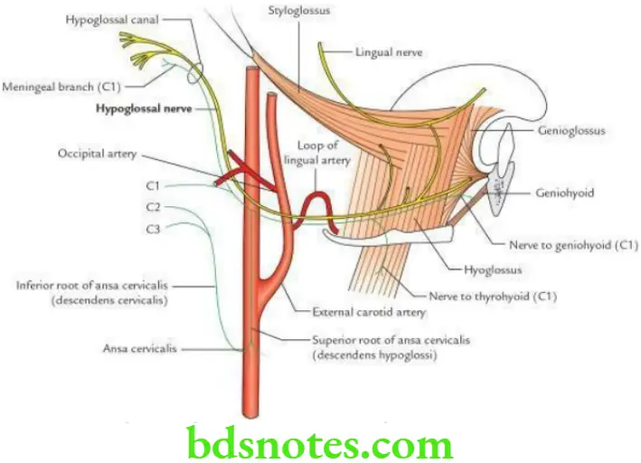
Hypoglossal Nerve Course and Relations The hypoglossal nerve arises from the hypoglossal nucleus located in the upper part of the medulla oblongata. It emerges from the anterior surface of the medulla between the olive and pyramid as 10–15 rootlets.
The fibres run anterolaterally and leave the posterior cranial fossa through the hypoglossal canal (anterior condylar canal). After emerging from the skull, it runs vertically downwards between the internal jugular vein and the internal carotid artery.
“Importance of studying the hypoglossal nerve for medical students: Questions explained”
“Common challenges in understanding hypoglossal nerve anatomy effectively: FAQs provided”
At the lower border of the digastric (i.e. at the level of the angle of the mandible), the nerve curves forward horizontally, crossing in front of internal and external carotid arteries, hooking around the origin of the occipital artery, crossing in front of the loop of lingual artery, and then runs on the superficial surface of hyoglossus. At the anterior border of the hyoglossus muscle, it enters the genioglossus and breaks up into terminal branches.
Hypoglossal Nerve Function
Hypoglossal Nerve Branches and distribution
“Steps to explain the anatomy of the hypoglossal nerve: Origin vs pathway vs innervation: Q&A guide”
- Branches of the hypoglossal nerve proper: They supply all the muscles of the tongue (intrinsic and extrinsic) except palatoglossus, which is supplied by the cranial root of the accessory via the pharyngeal plexus.
- Branches of hypoglossal nerve containing C1 fibres:
- The ventral ramus of the 1st cervical nerve, C1 joins the hypoglossal nerve below the skull. The fibres of C1 are distributed through the following branches of the hypoglossal nerve as follows:
- Meningeal branch
- Descendants hypoglossal/superior root of ansa cervical
- Nerve to thyrohyoid
- Nerve to geniohyoid
“Factors influencing success with hypoglossal nerve knowledge: Q&A”
Hypoglossal Nerve Applied anatomy The lesion of the hypoglossal nerve leads to paralysis of all the muscles of the tongue on the side of the lesion. This leads to deviation of the tongue on the side of the lesion protruding the tongue.
Hypoglossal Nerve Clinical Testing: The hypoglossal nerve is tested clinically by asking the patient to protrude his/her tongue.
In the lesion of the hypoglossal nerve, the protruded tongue deviates to the same side, i.e. side of the lesion. Thus, the deviated position of the protruded tongue indicates the side of the lesion.
Leave a Reply