• Skip to main content
  • Skip to secondary menu
  • Skip to primary sidebar
  • About Us
  • Terms of Use
  • Privacy Policy
  • Disclaimer
  • Contact Us
  • Sitemap

BDS Notes

BDS notes, Question and Answers

  • Public Health Dentistry
  • Periodontics
  • Pharmacology
  • Pathology
  • Orthodontics
    • Anchorage In Orthodontics
    • Mandibular Growth, Functional Matrix
    • Retention and Relapse
  • General Surgery
    • Cysts: Types, Causes, Symptoms
    • Maxillofacial Fractures, Disorders, and Treatments
    • Lymphatic Disorders
    • Neurological and Facial Disorders
  • Temporal And Infratemporal Regions
    • Spinal and Neuroanatomy
  • Dental Materials
    • Dental Amalgam
Home » Cerebrospinal Fluid Flow: Anatomy And Functions

Cerebrospinal Fluid Flow: Anatomy And Functions

February 7, 2025 by Kristensmith Taylor Leave a Comment

Cerebrospinal Fluid Flow: Anatomy And Functions

Describe subarachnoid cisterns in brief.

Answer. The subarachnoid space is the space between the arachnoid mater and the pia mater. It surrounds the CNS. The subarachnoid space around the brain is continuous with the subarachnoid space around the spinal cord. In the region of the brain, it communicates with the ventricular system of the brain.

Subarachnoid Cisterns

In certain situations, the subarachnoid space around the brain enlarges to form pools of cerebrospinal fluid (CSF) called subarachnoid cisterns.

The principal subarachnoid cisterns:

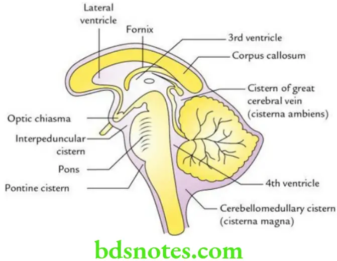
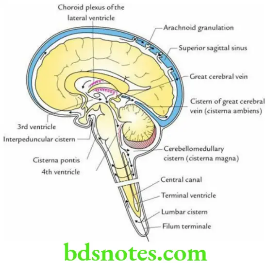
Cerebellomedullary Cistern: It is the largest cistern (also called cisterna magna) and lies in a triangle formed by the cerebellum, medulla oblongata, and occipital bone. It receives the CSF from the 4th ventricle through a foramen Magendie and foramina of Luschka.

Pontine Cistern: It lies on the ventral aspect of the pons and contains a basilar artery.

Interpeduncular Cistern (basal cistern): It lies in the region of the interpeduncular fossa and contains a circle of Willis.

Cistern Of Lateral Sulcus (Sylvian cistern): It occupies the lateral sulcus and contains the middle cerebral artery.

Cistern Of Great Cerebral Vein (cisterna ambient): It occupies the interval between the splenium of the corpus callosum and the superior surface of the cerebellum.

Head And Neck Meninges and cerebrospinal fluid Sagittal section of the brain showing location of principal subarachnoid cisterns

Head And Neck Meninges and cerebrospinal fluid Circulation of cerebrospinal fluid from its sites of formation

Filed Under: Head And Neck

Reader Interactions

Leave a Reply Cancel reply

Your email address will not be published. Required fields are marked *

Primary Sidebar

Recent Posts

  • Anodontia: Understanding the Congenital Absence of Teeth
  • Amelogenesis Imperfecta
  • Mottled Enamel: Understanding Fluoride-Induced Hypoplasia
  • Anomalies Affecting The Number Of Teeth
  • Supernumerary Teeth: Classification, Complications, and Care
  • Lingual Thyroid: When the Thyroid Grows on the Tongue
  • Developmental Anomalies Of The Tongue
  • Dentin Dysplasia: Understanding the Rootless Tooth Disorder
  • Median Rhomboid Glossitis
  • Histopathological Features Of Shell Teeth
  • Mesiodens And Its Impact On Eruption And Alignment
  • Microdontia: When Teeth Are Smaller Than Normal
  • Taurodontism: Understanding the Bull-Shaped Tooth Anomaly
  • Gemination vs Fusion: Developmental Dental Anomalies Explained
  • Tooth Anomalies Explained: From Size and Shape to Structure
  • Understanding Environmental Enamel Hypoplasia
  • Aplastic Anemia: Causes, Clinical Clues, and Treatment
  • Sickle Cell Anemia: A Clinical and Dental Perspective
  • Understanding Purpura and Its Oral Clues
  • Fluorosis Symptoms, Causes And Treatment

Copyright © 2025 · Magazine Pro on Genesis Framework · WordPress · Log in