Cavernous Sinus
Describe the Cavernous Sinus under the following headings:
- Cavernous Sinus Formation and location,
- Cavernous Sinus Relations,
- Cavernous Sinus Contents,
- Cavernous Sinus Tributaries and communications and
- Cavernous Sinus Applied anatomy.
Answer.
Cavernous Sinus Formation And Location
“Understanding the anatomy of the cavernous sinus through FAQs: Q&A explained”
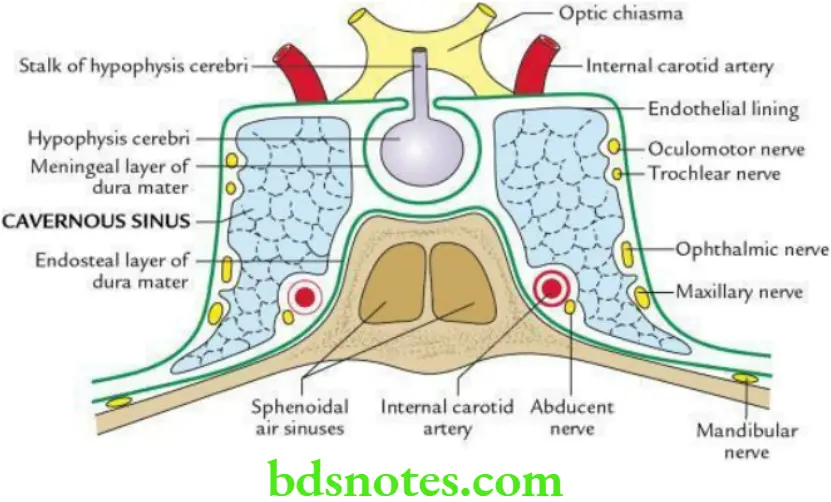
The two cavernous sinuses are situated on either side of the pituitary fossa and the body of the sphenoid. Each cavernous is a large venous space (2 cm long and 1 cm wide) formed by the separation of endosteal and meningeal layers of dura mater, lined by endothelium. Its floor is formed by the endosteal layer, whereas its lateral wall, roof and medial wall are formed by a meningeal layer of the dura mater.
“Importance of studying the cavernous sinus for medical students: Questions explained”
Cavernous Sinus Relations
Cavernous Sinus Superior: Optic chiasma, internal carotid artery and anterior perforated substance.
Cavernous Sinus Inferior: Foramen lacerum and greater wing of sphenoid.
Cavernous Sinus Medial: Hypophysis cerebri and sphenoidal air sinus.
Cavernous Sinus Lateral: Temporal lobe of the brain (uncus) and cavum trigeminal with trigeminal ganglion within it.
Cavernous Sinus Contents
Structures present in the lateral wall: From anterior to posterior, these are
- Oculomotor nerve (CN 3)
- Trochlear nerve (CN 4)
- Ophthalmic nerve (CN V1)
- Maxillary nerve (CN V2)
“Common challenges in understanding cavernous sinus anatomy effectively: FAQs provided”
Cavernous Sinus Structures Passing Through The Sinus
- An internal carotid artery with a sympathetic plexus around it
- Abducent nerve – below and lateral to the internal carotid artery
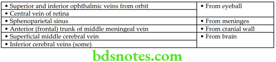
Cavernous Sinus Tributaries
“Factors influencing success with cavernous sinus knowledge: Q&A”
“Steps to explain cavernous sinus anatomy: Boundaries vs contents vs venous drainage: Q&A guide”
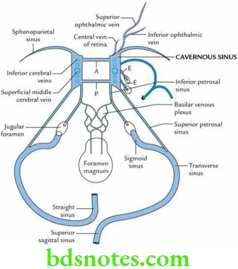
Cavernous Sinus Communications
- The superior petrosal sinus connects it with the transverse sinus.
- The inferior petrosal sinus connects it with the internal jugular vein.
- Emissary veins connect it with the pterygoid venous plexus.
- The ophthalmic vein connects it with the facial vein.
- Anterior and posterior intercavernous sinuses connect it with the opposite cavernous sinus.
“Role of cranial nerves in the cavernous sinus: Questions answered”
Cavernous Sinus Applied Anatomy
- Thrombosis of Cavernous Sinus: It may be caused by the spread of septic infection in the dangerous area of the face.
The clinical features of cavernous sinus thrombosis are as follows:- Severe pain in the eye due to involvement of the ophthalmic nerve.
- Ophthalmoplegia due to involvement of CN 3, 4 and 6.
- Oedema of eyelids due to congestion of orbital veins.
- Exophthalmos due to congestion of orbital veins.
“Early warning signs of gaps in understanding cavernous sinus basics: Common questions”
- Arteriovenous Fistula: It is caused by a rupture of the internal carotid artery into the cavernous sinus. It results in:
- Unilateral pulsating exophthalmos
- Loud systolic thrill/murmur over the eye
Leave a Reply