Axillary Nerve: Branches, Applied Anatomy
“What is the axillary nerve? A detailed question and answers guide”
- Axillary nerve is one of the two large terminal branches of the posterior cord of branchial plexus
Axillary nerve Course:
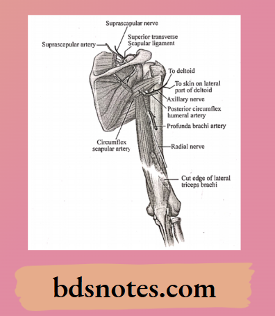
- From its origin, it passes backwards between subscapularis & teres major through the quadrangular space
- Here it lies in contact with the surgical neck of the humerus, just below the capsule of the shoulder joint
“Understanding the axillary nerve through FAQs: Branches, innervation, and applied anatomy explained”

“Importance of studying the axillary nerve for anatomy students: Questions explained”
Read And Learn More: BDS Previous Examination Question And Answers
Axillary nerve Branches:
- Muscular branchsupply deltoid & teres minor muscles
- Articular branch-supply shoulder joint
- Cutaneous branchsupply upper lateral cutaneous nerve of arm
- Branch to the shoulder joint which is divides into
- Anterior branch
- Supplies deltoid, pierces muscle & reaches skin
- Posterior branch
- Motor nerve to teres minor
- Cutaneous nerve to the arm
- Supplies posterior fibres of deltoid
- Anterior branch
“Common challenges in mastering axillary nerve notes effectively: FAQs provided”
Axillary nerve Applied Anatomy:
- The nerve may be injured during downward dislocation of shoulder joint
- It may be injured during fracture of surgical neck of humerus
- Injury to the nerve paralyses the deltoid muscle causing flattening of shoulder
- Wrongly injected drugs like penicillin may cause paralysis of the nerve
“Factors influencing success with axillary nerve studies: Q&A”
Question 2. Spinal Duramater.
Answer:
- It is a thick, tough fibrous membrane which forms a loose sheath around the spinal cord
- It is continuous with the meningeal layer of the cerebral duramater
“Steps to explain clinical relevance of the axillary nerve: Nerve injuries vs shoulder dislocations: Q&A guide”
Spinal Duramater Extend:
- From the foramen magnum to the lower border of the second sacral vertebrae
Spinal Duramater Significance:
- It gives tubular prolongations to the dorsal & ventral nerve roots & to the spinal nerves as they pass through the intervertebral foramina
Leave a Reply