• Skip to main content
  • Skip to secondary menu
  • Skip to primary sidebar
  • About Us
  • Terms of Use
  • Privacy Policy
  • Disclaimer
  • Contact Us
  • Sitemap

BDS Notes

BDS notes, Question and Answers

  • Public Health Dentistry
  • Periodontics
  • Pharmacology
  • Pathology
  • Orthodontics
    • Anchorage In Orthodontics
    • Mandibular Growth, Functional Matrix
    • Retention and Relapse
  • General Surgery
    • Cysts: Types, Causes, Symptoms
    • Maxillofacial Fractures, Disorders, and Treatments
    • Lymphatic Disorders
    • Neurological and Facial Disorders
  • Temporal And Infratemporal Regions
    • Spinal and Neuroanatomy
  • Dental Materials
    • Dental Amalgam
Home » Amyloidosis

Amyloidosis

November 20, 2025 by Kristensmith Taylor Leave a Comment

Amyloidosis

Write a short note on Amyloidosis.
Or
Write a short answer on amyloidosis.
Answer:

Amyloidosis is the term used for a group of diseases characterized by the extracellular deposition of a fibrillar proteinaceous substance called amyloid.

Classification of Amyloidosis
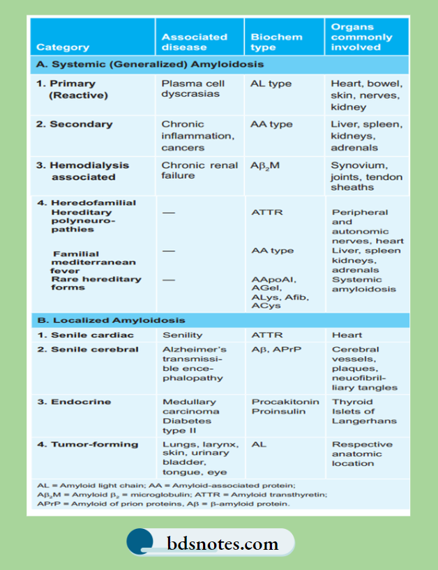
Including Amyloidosis-Classification-Of-Amyloidosis

Primary Amyloidosis

  • Biochemical Composition: AL protein
  • Associated Organ: Kidney, heart, bowel, nerve, and skin
  • Associated disease: Plasma cell dyscrasias, multiple myeloma, B-cell lymphoma.

Amyloidosis Pathogenesis

Morphology of Amyloidosis

  • Primary amyloidosis: This affects the kidney, liver, spleen, lymph node, adrenal gland, and thyroid gland
  • Secondary amyloidosis: This affects the heart, kidney, GIT, peripheral nerves, skin, and tongue.

Amyloidosis Gross Features

  • Affected organs are fim, enlarged, and waxy.
  • Painting the cut surface with iodine imparts yellow color which changes to bluish violet after the application of sulphuric acid.

Amyloidosis pathology

Pathologic Changes in Amyloidosis of Organ

  • The affected organ is usually enlarged, pale, and rubbery cut surface shows fim, waxy and translucent parenchyma.
  • The deposits of amyloid are found in extracellular locations, initially in the walls of small blood vessels producing microscopic changes and effects.

Filed Under: Pathology

Reader Interactions

Leave a Reply Cancel reply

Your email address will not be published. Required fields are marked *

Primary Sidebar

Recent Posts

  • Branchial Cleft Cyst: Background, Pathophysiology, Etiology
  • Maxillary Nerve: Origin, Course, And Branches
  • The Father Of Anatomy And A Great Anatomist Herophilus
  • Bone Structure – Anatomy
  • The External Carotid Artery: Anatomy, Branches, And Functions
  • Occipitofrontalis Muscle
  • Superficial Temporal Artery
  • Platysma Muscle
  • Cartilage
  • Cauda Equina And Conus Medullaris Syndromes
  • Subcutaneous Injections And Device Management
  • Types Of Circulation: Pulmonary, Systemic, And Portal
  • Structure Of Skeletal Muscle
  • Elastic Cartilage
  • Cellular Organelles And Structure
  • The Golgi Apparatus – The Cell
  • The Cytoplasmic Inclusions Of Certain Plant Cells
  • Dental Abscess
  • Laser Surgery
  • Our Facial Muscles And Their Functions

Copyright © 2026 · Magazine Pro on Genesis Framework · WordPress · Log in