Development And Growth Of teeth
Question 1. Describe the detail about stages of tooth development.
Answer:
Stages of tooth development:
- The development of a tooth is divided into several stages that are named after the shape of the enamel organ.
“Understanding tooth growth through FAQs: Stages, functions, and uses explained”
1. Bud stage:

“Importance of studying tooth growth for dental students: Questions explained”
Read And Learn More: BDS Previous Examination Question And Answers
- The epithelium of the dental laminae is separated from the underlying ectomesenchyme by a basement membrane.
- Round or avoid swellings develop from the basement membrane of 10 different points corresponding to tire 10 deciduous teeth.
- These are the primordial of the enamel organs, the tooth buds.
- In the bud stage, the enamel organ consists of peripherally located low columnar cells and centrally located polygonal cells.
- The supporting ectomesenchymal cells are packed closely beneath and around the epithelial bud.
- These cells undergo mitosis.
- As a result of it and the migration of the neural crest cells into it condensation of the tooth bud occurs.
- Condensation is immediately subjacent to the enamel organ in dental papillae.
- It forms tooth pulp and dentin.
- Similarly, ectomesenchymal condensation around tooth buds and dental papillae called dental sacs forms cementum arid periodontal ligament.
“Common challenges in mastering tooth growth notes effectively: FAQs provided”

“Why is proper understanding of tooth growth critical for diagnosing dental disorders? Answered”
2. Bud to cap transition:
- The transition from bud to cap marks the onset of morphologic differences between tooth germs that give rise to different types of teeth.
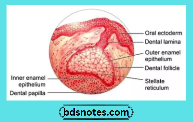
3. Cap stage:
- As the tooth bud continues to proliferate, due to unequal growth in different parts of the tooth bud, it leads to the cap stage.
- The peripheral cells covering the convexity of the cap are cuboidal called the outer enamel epithelium.
- The cells in the convexity of the cap called the inner enamel epithelium are tall, and columnar.
- Between these two layers are star-shaped cells called stellate reticulum.
- The outer enamel epithelium is separated from the dental sac and the inner enamel epithelium from the dental papilla by a basement membrane.
- The inner enamel epithelium meets the outer enamel epithelium at the rim of the enamel organ called the cervical loop.
“Factors influencing success with tooth growth studies: Q&A”
4. Bell stage:
- The continued growth of tire tooth germ leads to the bell stage as the tire enamel organ resembles a bell as the undersurface of the epithelial cap deepens.
- During the tills stage, the tooth crown assumes its final shape.
- Four different types of epithelial cells can be identified.
- Inner enamel epithelium:
- Consists of a single layer of cells of 4 – 5 pm in diameter and about 40 um high.
- These cells differentiate into ameloblast.
- Stratum intermedium
- It is essential for enamel formation.
- Cells in this layer are closely attached by desmosomes.
- Stellate reticulum
- It consists of star-shaped cells which help for attachment between the cells.
- It collapses to reduce the distance between the centrally situated ameloblasts and the nutrient capillaries near the outer enamel epithelium.
- Outer enamel epithelium
- The cells of this layer are cuboidal.
- Its smooth surface is laid down in folds between which capillary loops provide a rich nutritional supply for intense metabolic activity.
- Before the inner enamel epithelium begins to produce enamel, the peripheral cells of dental papilla differentiate into odontoblasts forming dentin.
- Remnants of dental lamina
- Outer enamel epithelium
- Ameloblasts
- Stellate reticulum Dental follicle Stratum intermedium Odontoblasts
- Collasped stellate reticulum Ameloblasts
- Inner enamel epithelium:
“Steps to explain stages of tooth development: Bud vs cap vs bell: Q&A guide”

“Role of the bud stage in initiating tooth formation: Questions answered”
5. Advanced bell stage:
- It is characterized by mineralization and root formation.
- The boundary between inner enamel epithelium and odontoblasts outlines the dentin enamel junction (DEJ).
- the dentin formation begins at this point and then proceeds pulpal and apically.
- Over it, ameloblast by down enamel which then proceeds coronally and cervically.
- In the cervical portion, the enamel organ gives rise to the epithelial root sheath of Hertwig.
“Differential applications of traditional vs modern teaching tools: Questions answered”
Question 2. Enumerate stages of tooth development. Describe the bell stage in detail.
Answer:
Stages of tooth development:
- Bud stage
- Cap stages
- Bell stage
- Advanced bell stage.
Leave a Reply