Pyogenic Granuloma
- Pyogenic granuloma is considered as an exaggerated conditioned response to minor trauma.
- Pyogenic granuloma is a misnomer since condition is not associated with pus formation.
“Understanding The Role Of Trauma In Causing Pyogenic Granuloma”
Pyogenic Granuloma Etiology
- It is caused by microorganisms such as streptococci and staphylococci.
- If there is minor trauma to the tissue it provides the pathway for the non-specifi microorganisms which can cause pyogenic granuloma.
- Hormonal imbalance can lead to pyogenic granuloma.
- The sulfhdryl molecule is the agent which lead to pyogenic granuloma.’
pyogenic granuloma
“Importance Of Early Detection Of Pyogenic Granuloma”
Pyogenic Granuloma Clinical Features
- It occurs at the age of 10 to 40 years.
- Female predilection is present.
- Most affcted sits are lip, gingiva, tongue, palate, vestibule.
- Lesion is more common in maxillary anterior region.
- Lesion is elevated, pedunculated or sessile mass with a smooth, lobulated or warty surface which is ulcerated.
- On manipulation, the ulcer bleeds.
- Lesion is pink to red to purple in color depending on age of the lesion. It is usually painless and is soft in consistency.
- Size of the lesion ranges from 1 mm to centimeters.
oral pyogenic granuloma
“Early Signs Of Pyogenic Granuloma On The Skin”
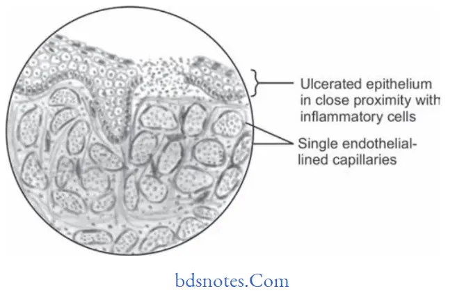
Pyogenic Granuloma Histopathology
- Overlying epithelium is thin and atrophic. At times, it is hyperplastic too.
- Surface of the epithelium is usually ulcerated and is replaced by thick firinopurulent membrane.
- Underlying connective tissue has number of endothelial lined vascular spaces engorged with RBCs and extreme proliferation of firoblasts and budding endothelial cells.
- There is presence of moderate infitration of PMN leukocytes, lymphocytes and plasma cells.
- Areas of hemorrhage and hemosiderin pigmentation is seen in connective tissue stroma.
“Role Of Hormones In Causing Pyogenic Granuloma”
Pyogenic Granuloma Treatment
Surgical excision of the lesion is done.
Leave a Reply