What Causes Squamous Cell Carcinoma in the Mouth? Everything You Need to Know
Question 7. Write a short note on squamous cell carcinoma.
Answer:
Squamous cell carcinoma
Squamous cell carcinoma is the most common malignant epithelial tissue neoplasm of the oral cavity. It is mostly derived from stratified squamous epithelium.
Squamous cell carcinoma Etiology
The following are the etiological factors that lead to squamous cell carcinoma:
“Understanding oral squamous cell carcinoma: Causes and symptoms”
- Tobacco smoking: Cigarettes, Bidis, Pipes, Cigars, and Reverse smoking.
- Use of smokeless tobacco: Snuffipping, Gutkha, Tobacco chewing, Tobacco as a toothpaste.
- Alcohol: Drinking spirits, Drinking wines, Drinking beers
- Diet and nutrition: Vitamin A, B-complex, and C deficiency, Nutritional deficiency with alcoholism.
- Dental factors: Chronic irritation from broken teeth, 3 fitting or broken prostheses.
- Radiations: Actinic radiation, X-ray radiation
- Viral infections: Herpes simplex virus (HSV), Human papilloma virus (HPV), HIV, Epstein-Barr virus (EBV)
- Chronic infections: Candidiasis, Syphilis
- Genetic factors: Oncogenes, Tumor suppressor
- PreExisting oral diseases: Lichen planus, Plummer Vinson syndrome, discoid lupus erythematosus, OSMF.
Oral squamous cell carcinoma
“Importance of early detection of oral squamous cell carcinoma”
Squamous cell carcinoma Gross Features
Grossly squamous cell carcinoma has two features, i.e.
- More commonly an ulcerated growth with elevated growth and indurated margin is seen.
- Less often a raised fungating or polypoid verrucous lesion without ulceration is found.
What causes squamous cell carcinoma
“Impact of alcohol consumption on mouth cancer risk”
Squamous cell carcinoma Clinical Features
- Carcinomas mostly occur in the 4th to 7th decades of life.
- Males are more commonly affected
- The lower lip is the most common site, the second most common site is the lateral border of the tongue. Among all intraoral sites, the dorsum of the tongue and hard palate are the least common sites for oral squamous cell carcinoma.
- The initial lesion may be asymptomatic or can be presented as a white or red nodule or fissure over the oral mucosa.
- Initially, the lesion is usually painless.
- More advanced lesions present either as a fast enlarging, exophytic or invasive ulcer or sometimes as a large tumor mass or a verrucous growth.
- The ulcerated lesion often shows persistent induration around the periphery with an elevated and everted margin.
“Treatment options for squamous cell carcinoma in the mouth”
Squamous cell carcinoma Histological Features
As per the histological grading by Broder’s Classification of Oral Squamous Cell Carcinoma.
Well-Differentiated Squamous Cell Carcinoma
- Most of the squamous cell carcinomas histologically belong to the well-differentiated category.
- In this lesson, the tumor epithelial cells to a large extent resemble the cells of the squamous epithelium both structurally and functionally.
“Role of surgery in treating oral squamous cell carcinoma”
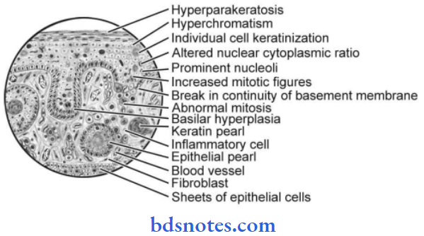
- Tumor cells produce large amounts of keratin in the form of “keratin pearls”.
- Tumor cells invade the underlying connective tissue, where the cells proliferate further and give rise to the formation of many epithelial islands within the connective tissue stroma
- Tumor cells often exhibit dysplastic features like cellular pleomorphism, nuclear hyperchromatism, individual cell keratinization, altered nuclear-cytoplasmic ratio, loss of cohesion, etc.
“Techniques for managing mouth cancer symptoms”
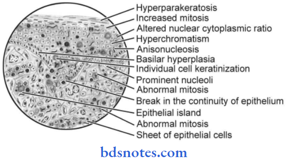
Moderately-Differentiated Squamous Cell Carcinoma
- The tumor cells are usually more severely dysplastic than that of the well-differentiated type.
- Tumor cells produce little or no keratin and these cells exhibit a greater number of mitotic cell divisions.
- There is the formation of epithelial islands or cell nests, etc. is diminished since these tumor cells do not differentiate or mature as much as the well-differentiated type of cells do.
“Pathophysiology of oral squamous cell carcinoma explained”
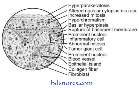
Poorly-Differentiated Squamous Cell Carcinoma
- In poorly differentiated squamous cell carcinoma, the malignant tumor cells produce no keratin.
- The tumor exhibits extensive cellular abnormalities with a lack of normal architectural patterns and loss of intercellular bridges between the tumor cells.
- Mitotic cell division is extremely high and because of this, the neoplastic cells are often very immature and primitive looking and it is often very difficult even to recognize them as squamous epithelial cells.
“Global prevalence of oral squamous cell carcinoma”
Leave a Reply