What Causes Dental Caries and How Is It Diagnosed
Dental Caries
Write a short note on dental caries.
Answer:
Dental caries
Dental caries is an “irreversible progressive microbial disease of the calcified tissues of the teeth, characterized by the demineralization of the inorganic portion and distortion of the organic substances of the tooth, which often leads to cavitation”.
Dental caries Etiopathogenesis
- Miller’s chemical-parasitic theory proposes that acid formed due to the fermentation of dietary carbohydrates by oral bacteria leads to progressive decalcification of the tooth. Structures with subsequent degeneration of the organic matrix.
- The acidogenic theory states that the process of dental caries involves two stages.
Dental caries
“Understanding dental caries: Causes and symptoms”
Initial Stage
Production of organic acid occurs as a result of the fermentation of carbohydrates by the plaque bacteria.
Later Stage
The acid causes decalcification of enamel followed by dentin and thereby causes total destruction of these two along with a dissolution of their softened residues. The final result is cavity formation.
“Impact of sugary diets on dental caries development”
Dental caries Gross Features
- The earliest change is the appearance of a small chalky white spot on the enamel which subsequently enlarges and often becomes yellow or brown and breaks down to form a carious cavity.
- The cavity becomes larger due to fractures of an enamel. As the lesion reaches the enamel-dentin junction, the destruction of dentin also begins.
Dental caries Histopathology
Stromal elements are present as loose connective tissue and as a myxoid, mucoid, and chondroid matrix which stimulates cartilage, i.e. pseudo cartilage. However true cartilage and bone are also observed in a small proportion of this tumor.
Causes of dental caries
“Radiographic features of advanced dental caries”
Histopathological Features of Caries in Enamel
Early Enamel Caries
- There will be loss of inter prismatic or inter rod substances with an increase in prominence of these enamel rods.
- The dark line often appears at the right angles of the enamel rods, suggesting segments.
- Accentuation of the incremental striae of Retzus often occurs.
“Case studies on outcomes of dental caries treatment”
“Complications of delaying dental caries diagnosis”
Advanced Enamel Caries
It presents several zones in the tissues, out of which four zones are clearly visible, starting from the inner advancing front of the lesion the zones are:
Zone 1: Translucent Zone
- It is the deepest zone that lies at the advancing front of the enamel lesion.
- This zone is more porous than normal enamel.
- The pores are larger than the normal enamel.
- The pore volume is 1%.
- This zone appears structureless.
- This zone contains more fluoride than normal enamel.
“Asymptomatic vs symptomatic stages of dental caries”
Zone 2: Dark Zone
- The dark zone is located just superficial to the translucent zone and its dark appearance is due to the excessive demineralization of the enamel.
- The zone is narrower in rapidly advancing caries and it is wider in slowly advancing lesions.
- The zone contains 2 to 4% pore volume.
- The pores are larger than normal but smaller than those of the translucent zone.
- This zone reveals some degree of remineralization of carious lesions.
Tooth decay causes
Zone 3: Body of Leison
- The zone is situated between the dark zone and the surface layer of the enamel.
- It represents the area of greatest demineralization.
- The pore volume is 5 to 25%.
- This zone contains appetite crystals larger than those of normal enamel.
- The large crystals result from the reprecipitation of minerals dissolved from the deeper zone.
“Treatment options for dental caries in dentistry”
Zone 4: Surface Zone
The surface zone when examined by the polarizing light appears relatively unaffected, it may be due to the surface remineralization by the salivary mineral ions.
Histological features of caries in dentin/dentinal caries
Dentinal caries histologically present 5 zones in the carious region, which are:
Zone 1: Normal Dentin
- This zone represents the innermost layer of the carious dentin, here the dentinal tubules appear normal.
- There is evidence of fatty degeneration of the Tome’s process.
- No crystals in the lumen of the tubules.
- No bacteria in the tubules.
- Intertubular dentin has normal cross-banded collagen and normal dense apatite crystals.
Zone 2: Subtransparent Dentin
- This is the zone of dentinal sclerosis and is characterized by the deposition of very fie crystal structures within the dentinal tubules.
- The superficial layer shows area of demineralization and damage of the odontoblastic processes.
- No bacteria in the tubules.
- Dentin is capable of remineralization.
“Role of dental fillings in treating cavities”
Zone 3: Transparent Dentin
- This zone appears transparent and this is because of the decalcification of dentin.
- It is softer than normal dentin.
- No bacteria in tubules.
- Cross-banded intertubular collagen is still intact.
- This zone is capable of self-repair and remineralization.
Zone 4: Turbid Dentin
- This zone is marked by the widening and distortion of dentinal tubules, which are packed with microorganisms.
- There is very little amount of minerals in dentin, and denaturation of collagen fibers also takes place.
- The zone cannot undergo self-repair or remineralization.
- This zone must be removed before restoration.
“Global prevalence of dental caries in different populations”
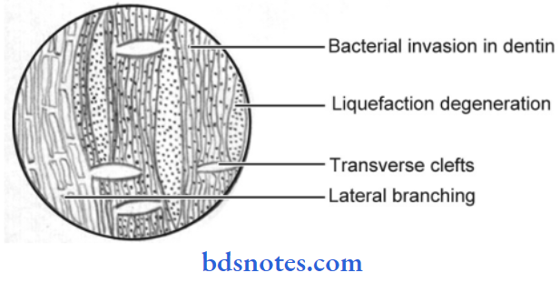
Zone 5: Infected Dentin
- This is the outermost zone of the carious dentin.
- It is characterized by complete destruction of dentinal tubules.
- In this zone, the area of decomposition of dentin, which occurs along the direction of dentinal tubules is called “Liquefaction foci of Miller”, which occur perpendicular to dentinal tubules and are called “Transverse Clefts”.
- In the process, the entire dentinal structure become destroyed and cavitation begins from the dentin enamel junction.
Dental caries Various Caries Activity Tests
Snyder Test
- This test measures the ability of salivary microorganisms to produce organic acids from carbohydrate metabolism.
- Glucose agar media containing an indicator dye, i.e. Bromocresol green is useful.
- The indicator dye changes from green to yellow in a range of pH between 5.4 to 3.8
- Paraffin-stimulated saliva is added into the medium, the change of the medium from green to yellow is indicative of the degree of caries activity.
“Follow-up care after dental caries treatment”
Salivary Reductase Test
- It measures the activity of the Reductase enzyme present in salivary bacteria.
- Paraffin-stimulated saliva is collected in the plastic container and an indicator dye “Diazoresorcinol” is added to it which colors the saliva blue.
- The Reductase enzyme liberated by the cariogenic bacteria causes color changes in the medium from blue to other colors, which indicates caries’ “conduciveness” of the patients.
Dental caries Alban’s Test
It is the modification of the Synder test. It uses less agar i.e. 5 mL per tube. The saliva is drooled directly into the tubes and the tubes are incubated for 4 days at 37°C. The color change is noted from bluish-green to yellow and the depth to which the change has occurred is noted.
“Emerging research on dental caries prevention”
Dental caries Strip Test for S. mutans Level in Saliva
Saliva/plaque samples are obtained by using tongue blades and toothpicks (after air drying the tooth for plaque samples) and are transferred to the S. mutans strip which is incubated in MSB agar (Mitis Salivarius Bacitracin agar).
The number of S mutant colonies is used to estimate the caries activity and more than 105 colonies per mL of saliva is indicative of high caries activity.
Dental caries Buffer Capacity Test
10 mL of stimulated saliva is collected at least once after eating and stored under paraffin oil to prevent the loss of volatile bicarbonate ions, 4 mL of this is measured in a beaker.
After correcting the pH meter to room temperature the pH of the saliva is adjusted to 7.0 by the addition of acid or base. The level of lactic acid in the graduated cylinder is then again recorded.
Lactic acid is then added to the sample until a pH of 6.0 is reached. The amount of lactic acid needed to reduce ph from 7.0 to 6.0 is the measure of the buffer capacity.
Leave a Reply