Vesiculobullous Lesions Of The Oral Cavity
Question. Enumerate vesiculobullous lesions and describe pemphigus.
Answer.
Pemphigus
Pemphigus is a group of vesiculobullous lesions of the skin and mucous membrane, which is characterized by the formation of intraepithelial vesicles or bulla, causing separation ofthe epithelium.
“Understanding vesiculobullous lesions through FAQs: Q&A explained”
Clinical Features Of Pemphigus
- It occurs during the 4th, 5th, and 6th decades of life and is more prevalent among females.
- Rapidly developing vesicle or bulla on several areas of skin and mucous membrane, which initially contain clear fluid, but later on, there is formation of pus.
- Vesicle ruptures very soon and leaves painful, erythematous ulcers that bleed profusely.
- Gentle traction or oblique pressure on and affected area around the lesion causes stripping of the normal skin or mucous membrane which is known as “Nikolsky’s Sign”.
- The patient may die due to dehydration and septicemia.
“Importance of studying vesiculobullous lesions for better diagnostic outcomes: Questions explained”
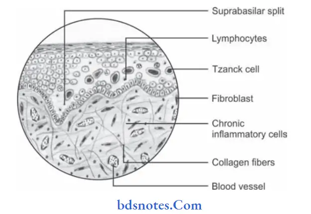
Histopathology Of Pemphigus
- Formation of a vesicle or bulla within the epithelium that results in the supra-basilar split.
- Following the suprabasal split the basal cell layer remains attached to lamina propria and appears as row of “Tomb Stone”.
- Loss of intracellular bridges and collection of edema flid results in acantholysis within spinous cell layer which causes disruption of prickle cell layer.
- As a result of acantholysis, clumps of large hyperchromatic epithelial cells lie free within the vesicular fluid; these desquamated cells are round and smooth in appearance and are known as Tzanck cells.
- A small number of PMNs and lymphocytes may be found.
“Common challenges in diagnosing vesiculobullous lesions effectively: FAQs provided”
“Steps to explain causes of vesiculobullous lesions: Autoimmune vs infectious factors: Q&A guide”
Treatment Of Pemphigus
- High dose of steroids
- Immunosuppressive agents
- Antibiotics to prevent secondary infection.
- Fluid and electrolyte balance must be strictly maintained.
Leave a Reply