Venous Diseases: Varicose Veins Of The Lower Leg
Write short note on varicose veins of lower leg.
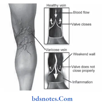
Answer. Dilated, tortuous, and elongated superficial veins of limb are called as varicose veins.
Varicose veins of lower leg Symptoms
- Majority of the patients come, with dilated veins in the leg.
They are minimal to start with and at the end of the day they are sufficiently large because of the venous engorgement. - Dragging pain in the leg or dull ache is due to heaviness.
- Night cramps occur due to change in the diameter of veins.
- Aching pain is relieved at night on taking rest or elevation of limbs.
- Sudden pain in the calf region with fever and enema of the ankle region suggests deep vein thrombosis.
- Patients can present with ulceration, eczema, dermatitis,bleeding, etc. Symptoms of pruritis/itching and skin thickening are also seen.
Varicose Veins of lower leg Signs
- Dilated veins are present in the medial aspect of leg and the knee. Sometimes they, are visible in the thigh also.
- Single dilated varix at SF junction is called saphena varix.
It is due to saccular dilatation of the upper end of long saphenous vein at the saphenous opening. - Veins are tortuous and dilated
- Ankle flare is a group of veins nearer the medial malleolus.
Varicose veins of lower leg Diagnosis
- Venous Doppler: With the patient standing, Doppler probe is placed at saphenofemoral junction and later wherever required.
Basically by hearing the changes in sound, venous flow, venous patency, venous reflx can be well identifid. - Duplex scan: It is a ultrasonographic Doppler imaging technique which along with direct visualization of veins,gives functional or anatomical information and also the color map.
- Venography: Before introduction of venous Doppler, venography is the common method of diagnosis.
- Plethysmography: This is a non-invasive method which measures the volume change in leg.
- Arm foot venous pressure: Foot pressure is not more than 4 mm Hg above arm pressure.
- Varicography: In this non-ionic, iso-osmolar, non-throm-bogenic contrast is injected directly in variceal vein to get detailed anatomical mapping ofvaricose veins.
This is used in recurrent varicose veins.
Varicose veins of lower leg surgical treatment
Trendelenburg’s operation:
- An inguinal incision is made, long saphenous vein identifid and the 3 tributaries are ligated.
- Long saphenous vein is ligated close to the femoral vein juxta femoral flsh ligation.
- An incision is given in front of the medial malleolus and long saphenous is isolated.
- The lower end is ligated and the vein incised.
- A long metallic stripper is introduced within the vein and brought out from the long saphenous vein in the inguinal incision.
- A metallic head is connected to the stripper and the vein is avulsed.
- Tight crepe bandage is applied, inguinal incision is sutured and the limb is elevated.
Subfascial ligation ofCocket and Dodd:
- In this operation, perforators are identified deep to deep fascia and they are ligated subfascial. This is indicated in cases of perforator incompetence with saphenofemoral competence. This is also done by an endoscope.
Leave a Reply