• Skip to main content
  • Skip to secondary menu
  • Skip to primary sidebar
  • About Us
  • Terms of Use
  • Privacy Policy
  • Disclaimer
  • Contact Us
  • Sitemap

BDS Notes

BDS notes, Question and Answers

  • Public Health Dentistry
  • Periodontics
  • Pharmacology
  • Pathology
  • Orthodontics
    • Anchorage In Orthodontics
    • Mandibular Growth, Functional Matrix
    • Retention and Relapse
  • General Surgery
    • Cysts: Types, Causes, Symptoms
    • Maxillofacial Fractures, Disorders, and Treatments
    • Lymphatic Disorders
    • Neurological and Facial Disorders
  • Temporal And Infratemporal Regions
    • Spinal and Neuroanatomy
  • Dental Materials
    • Dental Amalgam
Home » Understanding Action Potential: Ion Movements Made Simple

Understanding Action Potential: Ion Movements Made Simple

February 6, 2026 by Kristensmith Taylor Leave a Comment

Understanding Action Potential: Ion Movements Made Simple

Describe with the help of diagram the lonic basis of action potential.
Answer:

Action potential:

  1. Resting membrane potential
    • It occurs due to the movement of ions causing ionic imbalance across the cell
    • This imbalance is brought by
      • Sodium potassium pump
    • It actively transports Nat and K+ ions in opposite direction
    • By this pump, 3 Na+ ions are pumped out while 2 K+ ions are moved inside the cell using ATP
    • This results in more positivity outside than inside the cell
      • Selective permeability of membrane
    • Transport channels are selective for movement of specific ions
    • Channels for negatively charged substances remains closed as a result such ions remains inside the cell leading to more negativity inside than outside
  2. Action potential

Action Potential

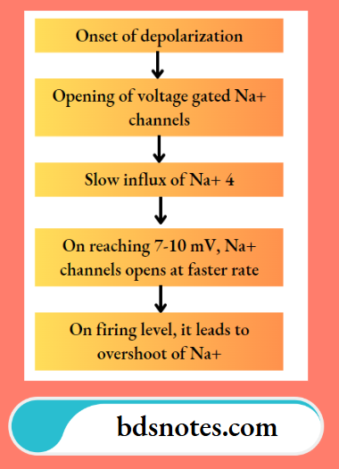
Similarly K+ channels opens and leads to efflux of K+ ions

Na+ channels are short lived while K+ channels remain open for longer duration

This results in efflux of more K+ ions producing more negativity inside the cell

Filed Under: Anatomy

Reader Interactions

Leave a Reply Cancel reply

Your email address will not be published. Required fields are marked *

Primary Sidebar

Recent Posts

  • Branchial Cleft Cyst: Background, Pathophysiology, Etiology
  • Maxillary Nerve: Origin, Course, And Branches
  • The Father Of Anatomy And A Great Anatomist Herophilus
  • Bone Structure – Anatomy
  • The External Carotid Artery: Anatomy, Branches, And Functions
  • Occipitofrontalis Muscle
  • Superficial Temporal Artery
  • Platysma Muscle
  • Cartilage
  • Cauda Equina And Conus Medullaris Syndromes
  • Subcutaneous Injections And Device Management
  • Types Of Circulation: Pulmonary, Systemic, And Portal
  • Structure Of Skeletal Muscle
  • Elastic Cartilage
  • Cellular Organelles And Structure
  • The Golgi Apparatus – The Cell
  • The Cytoplasmic Inclusions Of Certain Plant Cells
  • Dental Abscess
  • Laser Surgery
  • Our Facial Muscles And Their Functions

Copyright © 2026 · Magazine Pro on Genesis Framework · WordPress · Log in