• Skip to main content
  • Skip to secondary menu
  • Skip to primary sidebar
  • About Us
  • Terms of Use
  • Privacy Policy
  • Disclaimer
  • Contact Us
  • Sitemap

BDS Notes

BDS notes, Question and Answers

  • Public Health Dentistry
  • Periodontics
  • Pharmacology
  • Pathology
  • Orthodontics
    • Anchorage In Orthodontics
    • Mandibular Growth, Functional Matrix
    • Retention and Relapse
  • General Surgery
    • Cysts: Types, Causes, Symptoms
    • Maxillofacial Fractures, Disorders, and Treatments
    • Lymphatic Disorders
    • Neurological and Facial Disorders
  • Temporal And Infratemporal Regions
    • Spinal and Neuroanatomy
  • Dental Materials
    • Dental Amalgam
Home » Types Of Muscle Tissue: Striated, Non-Striated, Voluntary And Involuntary

Types Of Muscle Tissue: Striated, Non-Striated, Voluntary And Involuntary

February 6, 2026 by Kristensmith Taylor Leave a Comment

Types Of Muscle Tissue: Striated, Non-Striated, Voluntary And Involuntary

Question 1. Types of muscles.
Answer:

1. Depending upon the presence or absence of striations.

  1. Straited muscle.
    • These muscle contains cross striations.
    • Example: skeletal muscle, cardiac muscle.
  2. Nonstraited muscle.
    • They do not contains cross striation.

2. Depending upon action.

  1. Voluntary muscle.
    • The activities of it can be controlled at will.
    • Example: Skeletal muscle.
  2. Involuntary muscle.
    • The activites of it cannot be controlled at will.
    • Example: Cardiac smooth muscle.

3. Depending upon the location.

  1. Skeletal muscle.
    • Found in association with bone.
  2. Cardiac muscle.
    • Form musculature of heart.
  3. Smooth muscle.
    • Present in hallow viscera.

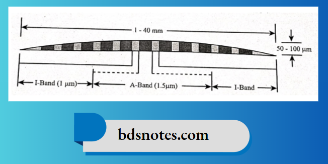
Question 2. Sarcomere.
Answer:

  • It is the structural and functional unit of the skeletal muscle.
  • The cross striations which are characteristic feature of skeletal muscle are due to alternate dark and light cross band.
    1. The dark line is highly refractive and is called ‘A’ band.
      • In the middle of `A’ band there is a light area called ‘H’ zone.
      • In the middle of ‘H’ zone lies the middle part of myosin filament called ‘M’ line.
    2. The light band is low refractive and is called ‘I’ band.
      • In the centre of each I band is found a narrow line of highly refractive material called ‘Z’ line.
    3. The substance included between two adjacent ‘Z’ line is called “Sarcomere”.


General Appearance

Filed Under: Anatomy

Reader Interactions

Leave a Reply Cancel reply

Your email address will not be published. Required fields are marked *

Primary Sidebar

Recent Posts

  • Branchial Cleft Cyst: Background, Pathophysiology, Etiology
  • Maxillary Nerve: Origin, Course, And Branches
  • The Father Of Anatomy And A Great Anatomist Herophilus
  • Bone Structure – Anatomy
  • The External Carotid Artery: Anatomy, Branches, And Functions
  • Occipitofrontalis Muscle
  • Superficial Temporal Artery
  • Platysma Muscle
  • Cartilage
  • Cauda Equina And Conus Medullaris Syndromes
  • Subcutaneous Injections And Device Management
  • Types Of Circulation: Pulmonary, Systemic, And Portal
  • Structure Of Skeletal Muscle
  • Elastic Cartilage
  • Cellular Organelles And Structure
  • The Golgi Apparatus – The Cell
  • The Cytoplasmic Inclusions Of Certain Plant Cells
  • Dental Abscess
  • Laser Surgery
  • Our Facial Muscles And Their Functions

Copyright © 2026 · Magazine Pro on Genesis Framework · WordPress · Log in